
MBI Graduate program: ME:120.754
Research & Thesis
Instructors: Corinne Sandone, Professor, Lead Instructor
David Rini, Professor
Jennifer Fairman, Professor
Juan Garcia, Professor
Lydia Gregg, Associate Professor
Sarah Poynton, Associate Professor
Jeffrey Day, Assistant Professor
Man-San Ma, Instructor
Credits: 12 credit hours:
summary
Original investigation with expository illustrations (may include 3D digital or physical model, 2D or 3D animated sequence, video or immersive technology) completed under a University approved faculty preceptor and department faculty advisor. All work complies with University research, publication, copyright, and patent policy.
Course Description
Objectives
- Contribute to medical illustration through research and communicate effectively in written and illustrative form.
- Formulate a statement of purpose or hypothesis as it pertains to a topic in biomedical communication.
- Communicate, consult, and negotiate effectively with experts in a given area of research.
- Apply critical thinking, time management, and organizational skills in research.
- Investigate accumulative literature relative to the thesis topic.
- Utilize effective writing skills in correspondence, proposals, reports, and articles for publication.
- Value the need for accuracy, originality, creativity, and craftsmanship in producing quality work.
- Produce a written thesis according to the prescribed University format.
- Select the most appropriate form(s) for illustrative components and prepare all visual material in accordance with its reproduction requirements.
Schedule
1st Quarter: 1 Lecture hour and 3 Studio hours per week
2nd Quarter: 3 Lecture hours and 24 Studio hours per week
3rd Quarter: 3 Lecture hours and 20 Studio hours per week
Assignments
Each student-preceptor-advisor team arranges individually according to project. Every project culminates ina written thesis and oral presentation.
Resources
- Department, School of Medicine, specialty, University libraries, inter-library loan systems.
- Preceptor and Advisor expertise.
- Facilities, materials, and equipment provided by preceptor, department, and outside sources as needed.
- Scholarship awards, when available.
Student Evaluations
- 1:1 faculty-student interface
- Scheduled preceptor-student and preceptor-advisor meetings
- Discussion of student’s own work from media courses
- Preceptor’s letter of thesis acceptance
- Thesis presented, reviewed, and approved by the School of Medicine MA-PhD Committee
- Final grade
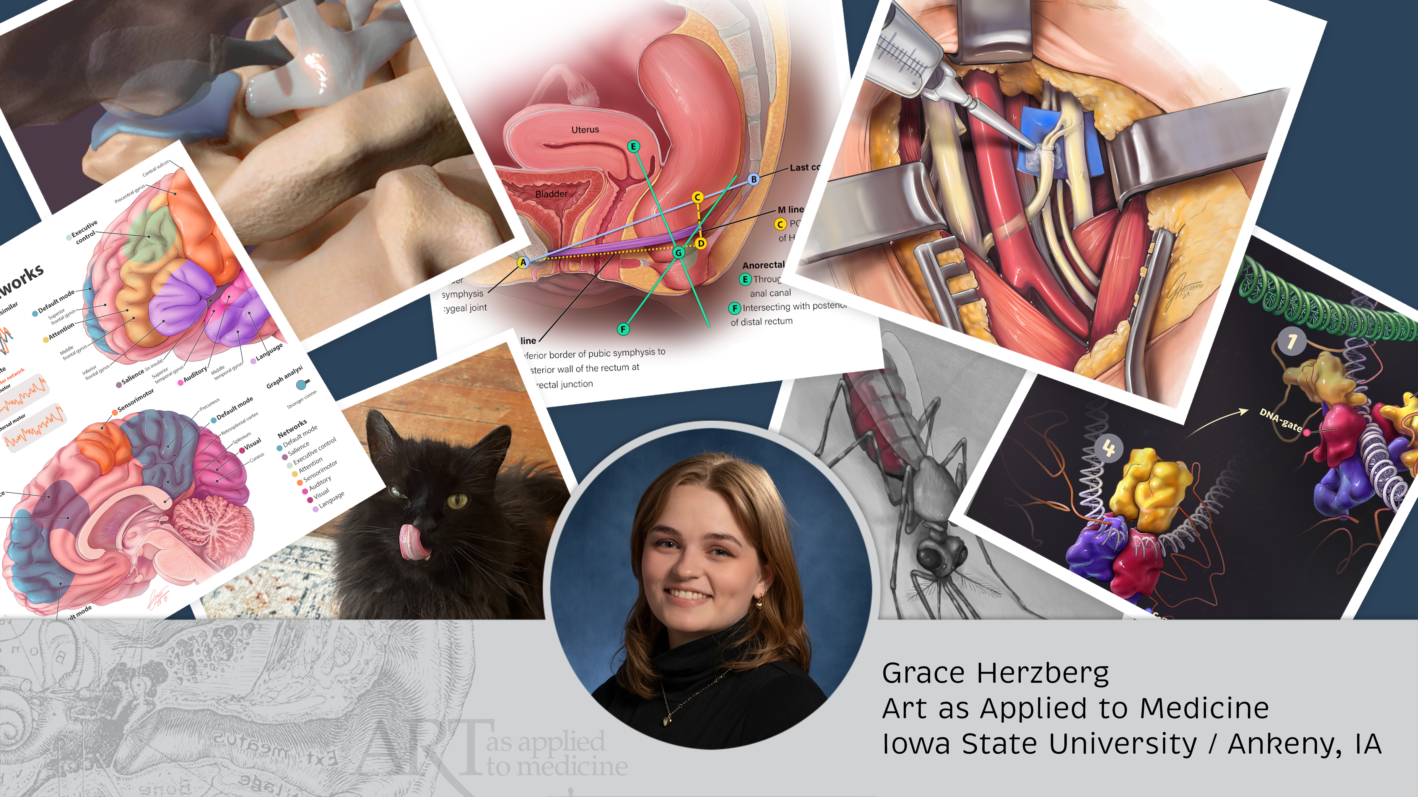
Student Artwork
Sample Thesis Projects
Gamifying Radiology Education: A Virtual Desk for Studying MRI Pulse Sequences
MRI physics, a challenging topic for radiology residents to master, is a key component of the American Board of Radiology (ABR) CORE exam required for board certification in diagnostic and interventional radiology. A previous needs assessment identified a desire for...

Development of A Sustainable Serious Game To Support Central Venous Catheter Placement Education
The central venous catheter kit, containing over 10 items, can overwhelm first-time trainees. To address this, we’re creating a learning experience that merges an online module with 3D-printed models for medical trainees to start identifying these items in the kit...
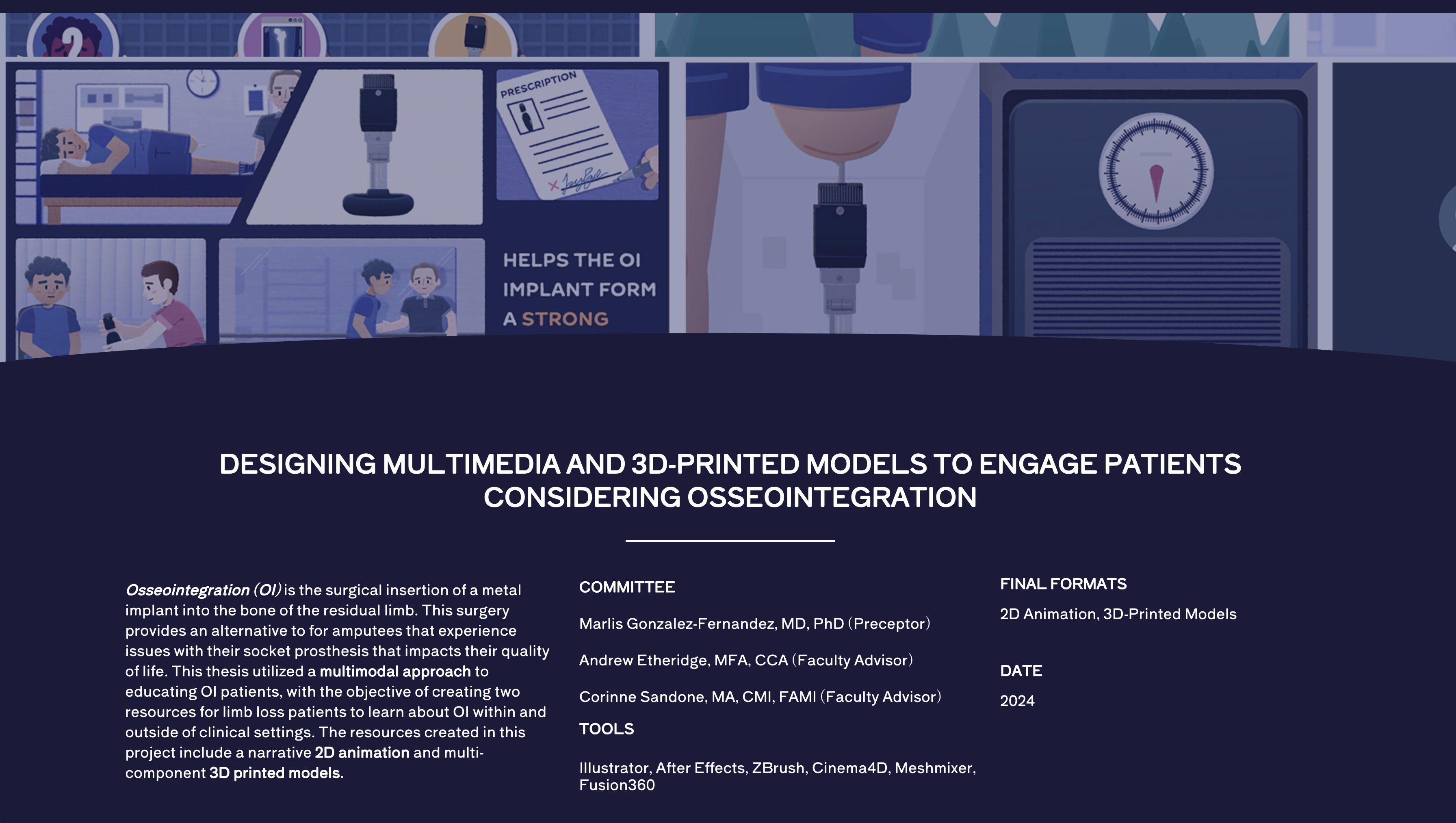
Designing Multimedia and 3D-Printed Models to Engage Patients Considering Osseointegration
Approximately 1 in 150 Americans currently live with limb loss and this number is expected to double by 2050 (Smidt and Bicknell 2024). While most lower limb amputees use a socket prosthesis to connect their residual limb to their prosthetic leg, some individuals...
Past Thesis Research
2024
From Parthanatos to PAANIB-1: A Multimedia Exploration of Cell Death Mechanisms and Therapeutic Strategies for Parkinson’s Disease :: Sarrah Hussain
Recent discoveries in molecular biology have shed light on a potential therapeutic treatment for Parkinson’s disease (PD)—a progressive neurological disorder marked by the gradual loss of dopaminergic neurons in the brain. This treatment involves selectively intervening in the parthanatos pathway, a preventative cell death mechanism that normally helps restrict the cell-to-cell spread of disease. This cell death pathway is characterized by its distinctive breakdown of the cell’s DNA into fragments that escape from the nucleus into the cytoplasm prior to degrading.
In addition to parthanatos, the cGAS-STING pathway is another molecular mechanism that produces a rapid microglial immune response upon detection of cytosolic DNA fragments. Thus, the cGAS-STING pathway is linked to the elimination of infected cells by sensing and responding to foreign DNA, as well as self-DNA fragments created through parthanatos. In neurogenerative diseases such as PD, both pathways become excessively engaged and overactivated—perpetuating a cycle of widespread inflammation and cell death.
Traditionally, it has been believed that blocking the initial stages of parthanatos would be the most effective in preventing DNA fragmentation and cell death. However, targeting the early steps of parthanatos has drawbacks, as this inadvertently hinders essential DNA repair. Therefore, new research has shifted focus downstream to prevent parthanatos-induced DNA cleavage without interfering with important upstream DNA repair processes. In particular, Macrophage Migration Inhibitory Factor (MIF) nuclease has been identified as the critical molecule for initiating self-DNA cleavage in cells, thereby triggering the decisive step of parthanatos.
Exciting new research has unveiled PAANIB-1, a first-in-class MIF inhibitor that prevents this final step of parthanatos, thus also preventing the generation of DNA fragments that over-activate the cGAS-STING pathway. Targeting MIF using PAANIB-1 therefore shows promising potential in its ability to slow or prevent inflammation and neuronal damage in PD. As further testing for PAANIB-1 viability proceeds, it is crucial for researchers, students, donors, and patients to understand both PD pathogenesis and this novel, highly-targeted intervention strategy. I aim to transform these findings into a didactic, highly engaging 3D animation and interactive web module, catering to both scientific and lay audiences.
Thesis Student
Department Advisor
How Contractility Kits Can be Used to Fight Cancer :: Nicholas Kilner-Pontone
Mutations that alter the flexibility of the cell’s cortex can contribute to many diseases, including cancer development and metastasis. This property of cells is not well understood and is the focus of the Robinson Lab at Johns Hopkins University School of Medicine. Their research has led to the discovery of protein condensates known as Contractility Kits (CKs), which has provided insight into how cell cortices respond to mechanical stress and how this flexibility is regulated. The ability to reduce the flexibility of cancer cell cortices has shown promise in developing therapies that prevent metastasis. Currently, the Robinson Lab has two patents for targeted use of the compound 4-HAP, which has shown the ability to increase the concentration of nonmuscle myosin II, a CK component that accumulates in the cortex. The ability to make cancer cells more rigid has shown promise in fighting pancreatic and colorectal cancers as well as potentially others.
This research is at the forefront of molecular and cellular biology, and the creation of visuals is catching up. Until now, these processes have been described mostly through static figure illustrations and video clips based on protein concentrations as their structures and behaviors come into focus. However, the association of CKs with the cortex and the behaviors of individual proteins that comprise them are dynamic. What is missing are educational animations of how these proteins sense mechanical force, how they associate and dissociate from the cortex and how this behavior can be modified.
To address this gap in visual material, we created a short 3D animation to describe the proteins that form CKs, as well as what is currently known about the mechanical feedback loops that govern CKs, and finally how small molecular modulators such as 4-HAP can alter the flexibility of cell cortices. This animation will supplement Dr. Robinson’s lecture material and help support his lab’s efforts to get the resources needed for human trials. The video can be found on Dr. Robinson’s website and YouTube, where anyone can access it.
Thesis Student
Preceptor
Department Advisor
Why Shed the Shield? A Narrative-based Educational Campaign for Discontinuing Gonadal Shielding :: Ann Seliger
Gonadal shielding, or placing a protective barrier between patients and a source of radiation to reduce exposure to the gonads, has long been a standard practice in diagnostic imaging. However, advances in technology and new evidence regarding radiation exposure have led to shielding gradually being discontinued at medical centers nationwide. Radiology technologists (RTs) are responsible for implementing “no-shield” policies at these centers, but the legacy status of shielding and conflicting guidance from healthcare professionals complicate messaging around shielding.
We created a visual narrative to convey the scientific rationale behind no-shield policies in an educational campaign for RTs. We recruited 41 professional RTs employed at Johns Hopkins to assess the animation. Learning was measured through a pre- and post-test and engagement was measured with Likert-scale surveys. Open response questions were analyzed qualitatively for themes.
We performed paired sample t-tests to check for differences in pre- and post-test scores. Our analysis found that scores significantly increased after RTs watched the animation (p = 0.00009). In the engagement survey, RTs generally agreed with positive statements about the animation’s content (attention, understanding, enjoyment).
Our study provides insight into the benefits of incorporating narrative-based animation into medical education campaigns. The narrative animation produced for this study will be available online and potentially distributed to hospitals for RT training as they adopt no-shield policies.
Thesis Student
Department Advisor
Pig-to-Human Kidney Xenotransplantation: Designing the First Patient Education Visuals :: Chloe Woodin
Kidney transplantation is considered the most effective treatment for end-stage renal disease, however, there is a severe shortage of available kidneys. Nearly 90,000 people in the US are waiting for a kidney transplant, but only 25,000 kidney transplants occur annually. Each day, 17 people die waiting for an organ transplant. In response to the organ shortage, Johns Hopkins is at the forefront of research into pig-to-human kidney xenotransplantation, with human clinical trials expected to begin as early as 2025.
As clinical trials approach, it is critical to provide kidney transplant waitlist candidates with adequate education so they can consider xenotransplant, which is a novel, complex treatment option. To educate patients prior to clinical trials, Johns Hopkins plans to conduct a series of three one-hour, physician-led patient information sessions to educate kidney transplant waitlist candidates about xenotransplantation. A prototype of visual materials to be used during the first of three educational sessions was designed and created to accompany the following topics: 1) background on xenotransplantation, 2) immune system and xenotransplant rejection, 3) genetic modification, and 4) where pig kidneys come from and wrap up. Three types of visuals were designed: standalone images, sequential image series, and 2D animations. Ninety individual visual assets were created and arranged into slide layouts and storyboards. A draft slideshow with 130 color illustration and storyboard slides was developed and iterated upon based on formative feedback from transplant surgeons and patient educators.
The result of this thesis is a 34-slide PowerPoint presentation consisting of 14 standalone illustrated slides, four sequential image series, and three 60-second 2D animations. Four additional slides and one animation storyboard were created as supplemental material. These are the first visuals depicting kidney xenotransplantation designed specifically for kidney transplant waitlist candidates.
Future directions for this project include conducting an IRB-approved study to evaluate the effectiveness of these visuals for the target audience, expanding the prototype to include additional topic areas, and adapting these visuals for web and print. This project successfully created a framework for patient education that can be built upon as xenotransplantation moves into accepted clinical practice.

Thesis Student
Preceptor
Department Advisor
2024 Thesis Presentations
![]() The Thesis Presentations of the MBI Class of 2024 will be held on Tuesday, May 21st, 2024, 3:00-4:30 PM (EDT), both in-person in the Chevy Chase Auditorium and with a Live broadcast to the Department’s YouTube Page. The students will present to their faculty, preceptors and those able to join us live.
The Thesis Presentations of the MBI Class of 2024 will be held on Tuesday, May 21st, 2024, 3:00-4:30 PM (EDT), both in-person in the Chevy Chase Auditorium and with a Live broadcast to the Department’s YouTube Page. The students will present to their faculty, preceptors and those able to join us live.
Alumni, students, and the public are invited to participate through YouTube Live Chat. A Q&A with Audience questions and Typed questions from the YouTube Chat will be held at the end of each presentation.
Links to the YouTube Live event will be shared to the Department’s social media accounts: Facebook, LinkedIn, Instagram, and Twitter.
Thank you for taking a look at the research into the art of biomedical visualization from the MBI Class of 2024!

2023
Class of 2023 – Thesis Research
- Lilas Armstrong-Davies – Visualizing Brain Size Evolution
- Courtney Brendal – MRI Artifact Detection
- Hao Wen Choi – Endoscopic Endonasal Skull Base Surgery
- Anna Mai – Visualizing MIPSA
- Gyyoung Oh – Understanding the Cellular Mechanisms of Parkinson’s
- Elizabeth Siedell – Illustrating New Visual Guide for Central Line Placement
- Emily Simpson – Improving Simulation Task Training
- Miranda Stano – Personalized Visualization of Congenital Heart Disease
Visualizing brain size evolution in platyrrhine monkeys: a skull reconstruction, endocast extrapolation, and volume comparison of Cebupithecia sarmientoi :: Lilas Armstrong-Davies
In 2018, at the paleontological site La Venta, Colombia, the first complete monkey skull from the Middle Miocene epoch (11-13 MYA) was recovered. Paleontologists identified the skull as Cebupithecia sarmientoi, an extinct species of platyrrhine monkey whose skull was only previously known by teeth and mandible fragments. C. sarmientoi is most likely related to the Pitheciidae family of platyrrhine monkeys but has a noticeably smaller cranial cavity. This discrepancy indicates that C. sarmientoi had a smaller brain size than its extant relatives. To confirm the relative brain size, a digital 3D model of the fossil was reconstructed and a cast of the cranial cavity, or endocast, was created. The volume of the endocast was compared to 16 closely related living species of platyrrhine monkey using an RMA regression and the relative brain sizes were placed in evolutionary trees. This is the first study to attempt to extrapolate an endocast after a skull reconstruction and the workflow was assessed for future research. These visualizations of C. sarmientoi are the first of their kind and provide new insights on the rate and timing of brain size increase in platyrrhine monkey evolution. The visualizations will contribute to scientific publications on the C. sarmientoi discovery and enhance public understanding and interest in evolution and paleontology.

Thesis Student
Preceptor
Department Advisor
MRI Artifact Detection: Schematic Design in Perceptual Training for Radiological Board Exam Preparation :: Courtney Brendal
MRI physics and clinical comprehension of artifacts, or the spectrum of visual anomalies that can occur in all types of imaging modalities, are cornerstone subjects in the American Board of Radiology CORE Exam. Use of perceptual training is important in developing radiologists’ abilities to detect artifacts on images but is underutilized in radiology education resources. This thesis project outlines the creation and investigation of the novel use of representational schematic images of MRI artifacts for perceptual training. The results of this study inform the direction of a web-based, interactive learning resource that radiology residents and other learners would find useful and engaging to use.
We created an online Qualtrics survey and distributed it to medical students and radiology residents in the Johns Hopkins University School of Medicine (JHUSOM). Study participants were randomized to interact with only the schematic images, only the MRI images, or a combination of the two image types. There was no significant difference in participant pre/post-test performance and engagement survey feedback across all three test conditions, which suggests that studying schematic images could be equally as useful as studying from MRI images. Participants praised the resource for its use of concise and manageable visuals and explanations. Respondents suggested adding more MRI artifact image examples for additional reference and creating a glossary for radiology terminology that could accommodate beginner learners.
With these insights, we used the content from the Qualtrics pre-and-post-tests to design an interactive quiz using Unity. Additionally, we also incorporated our artifact image media into a downloadable PDF reference sheet and Anki deck for students to review for their CORE Exam study needs. These resources will be hosted on teamrads.com, JHUSOM’s open education radiology website.

Thesis Student
Preceptor
Department Advisor
Endoscopic Endonasal Skull Base Surgery for Pituitary Lesions: An AI-Assisted Creative Workflow to Develop an Animated Educational Resource for Patients and Physicians :: Hao Wen Gilbert Chen
Endoscopic endonasal skull base surgery (EESBS) is a complex surgical approach used to access and remove pituitary lesions and tumors through the anatomically complex sinonasal corridor. Many patients find it difficult to understand and conceptualize the nuanced anatomy, surgical technique, and the substantial postoperative side effect and symptom profile associated with this minimally invasive surgical procedure.
Effective perioperative counseling is challenging in the setting of time-constrained preoperative visits and by the abstract surgical technique in an anatomical region that is unfamiliar to most patients. The goal of this project was to create an interactive and accessible resource available for both patients and surgeons alike to help improve perioperative counseling and patient education. This research involved using an Artificial Intelligence assisted workflow to create a series of 2D animations that aim to explain the treatment process using engaging, educational, and easy-to-understand content. Animations were designed to provide patients with an immersive experience to help explain relevant anatomy as well as preoperative, surgical, and postoperative treatment phases. The resulting narrated, 2D “cut paper” style animations are a resource to assist physicians with patient communication and education needs. Given its modular design, this resource can be edited or updated to reflect changes in surgical techniques and can be adapted into interactive e-learning materials.
Recognizing that generative Artificial Intelligence (AI) reached public awareness in early 2023 as a powerful tool for generating new content including text, images, and audio from existing online data, this project aimed to incorporate this novel technology to enhance patient education over traditional approaches. With the implementation of an AI-assisted creative workflow, we utilized 1) an AI art generator for style inspiration, 2) an AI chatbot for assistance in script drafting, and 3) an AI voice synthesizer for audio narration. The research also addresses some of the potential risks and concerns associated with applying AI-assisted tools to patient education content creation. By implementing these new technologies in the creative workflow, this research provides valuable insights into when and how to appropriately apply AI technology while creating patient education content. The use of AI-assisted technologies in creating patient educational resources represents an exciting new development in the field of medical communication.


Thesis Student
Preceptor
VISUALIZING MIPSA: Exploring Web-based User Experience Design in Teaching and Promoting Molecular Technology :: Anna Mai
This thesis explores the potential of web-based user experience in teaching and promoting a novel molecular technology, Molecular Indexing of Proteins by Self Assembly (MIPSA), which is a scalable and low-cost solution for comprehensive antibody profiling. Despite its potential applications, there are no existing visuals that explain the complex science of MIPSA. To address this gap, a website was developed with animations, graphics, and concise content to educate and engage the target audience of researchers and investors in biotechnology.
The website is the first of its kind to explain and visualize the concept of MIPSA using multimedia and the multimedia learning theory. The website aims to deliver the didactic content in an engaging and comprehensible way by employing website design principles, multimedia and the multimedia learning theory. A focus group was held with researchers and students at Johns Hopkins to assess the effectiveness of the website, and results showed high ratings for overall effectiveness, navigation, content organization, and visual appeal. The project’s implications for biocommunication and the promotion of a new molecular technology are discussed, along with suggestions for future studies…

Thesis Student
Preceptor
Department Advisor
Understanding the Cellular Mechanisms of Parkinson’s Disease: An Interactive Learning Module :: Gyyoung Oh
As the second most common neurodegenerative disorder, Parkinson’s disease (PD) is still being thoroughly researched. The complex cellular mechanisms of PD are responsible for its many symptoms, such as tremors, difficulties with balance, and constipation. An immense amount of information about PD has been discovered, but much of it remains among the scientific research community. There is a need for resources to educate PD patients, caretakers, and students about specific reasons behind diagnoses and symptoms. In addition, existing resources also do not allow audiences to customize their learning process.
This project aims to provide an effective interactive learning module that allows users to choose the order and rate at which they absorb Information. In particular, it aims to teach users about the cellular mechanism of parthanatos that causes many of the symptoms of PD. Using 3D models and animations, it was developed as a WebGL (Web Graphics Library) web application created with Unity. Cinema4D was used to convert
molecular structure data into 3D models.
The goal of the interactive learning modules is to provide the first step in filling the gap in existing resources and educating all users about parthanatos, whether it is to supplement an existing knowledge background or start from the beginning. In future additions to the modules, other cellular pathways can be added to expand the educational content of the project.


Thesis Student
Department Advisor
Illustrating and Evaluating a New Visual Procedural Guide for Central Line Placement :: Elizabeth G. Siedell
Central venous catheterization, or central line, is a small catheter placed in a vein for long-term drug therapy or dialysis. It is a common procedure learned and performed across many medical fields, with over 5 million central lines inserted every year in the United States alone. However, improper insertion can be lethal, and complications affect patient quality of life, hospital days, and costs. Immediate complications, including vascular, cardiac, and pulmonary, are directly related to improper procedure technique. Proper central line procedural guidance and training optimizes patient safety and decreases complications.
Traditionally, most clinical procedural guides are text-heavy manuals with few diagrams, which can be difficult to learn from or referenced during a procedure. In this project, we aimed to address this issue by creating a visual guide for venous catheterization so that medical trainees and practitioners can more safely and efficiently perform the procedure. We also created an IRB-approved study to compare text-only instruction against the novel visual guide through a simulated central line procedure in the Johns Hopkins Simulation Center. Using the two instructional methods, our study assessed time spent on procedure, accuracy of procedure, eye-tracking data, and postsurvey feedback.
We analyzed trends in the survey data to find differences in survey A (given to the users who used the text-only guide) and survey B (given to users who used the illustrated guide). The preliminary trends from the survey show reduced cognitive load and task load in participants who used the illustrated guide. Survey results also showed a trend in strong user preference for the illustrated guide compared to the text-only guide. Common feedback was in favor of the visual guide or expressing the need for visuals if they received the text-only guide. Our study suggests the importance of illustrated procedural guides in both user preference and in reducing user cognitive load. Moving forward, the study will continue to run and our findings could inform a better standard for creating healthcare procedural guides. Additionally, we plan to publish our research in a peer-reviewed journal to further advocate for the necessity of visual procedural guides.

Thesis Student
Preceptor
Improving Simulation Task Training: A Novel Interactive Resource and 3D Printing Provide Customizable Solutions for Increasing Fidelity and Lowering Costs :: Emily Simpson
Abscess incision and drainage (I & D) is a common procedure used for treating skin abscesses. This procedure can relieve pain and accelerate healing of an abscess; however, if performed inadequately, infection can spread. Medical simulation devices, called task trainers, permit learners to manually practice the skills and steps of a procedure. Abscess incision and drainage task trainers function as a bridge between book learning and practicing on a real patient.
Abscess task trainers are expensive; commercial trainers range from $30 – $150 per single-use device. Furthermore, they are available only in limited skin tone options. Limited exposure to skin tone and body mass diversity restricts adequate healthcare training. Simulation centers also may encounter supply chain issues when ordering abscess incision and drainage task trainers. To prevent this, a simulation center must order the trainers months in advance, however, some commercial trainers have a short shelf life. Instructions for several do-it-yourself alternatives exist, but the resulting task trainers are low fidelity and can be complicated to create.
To address these limitations, we developed a new method of abscess task trainer creation, including the design of a 3D printed base. The abscess task trainers created for this project are lower cost compared to commercial alternatives, and allow customizable fidelity, such as variety of skin tones, body mass, and the ability to add inflammation. To help simulation center facilitators navigate this customizability, we designed a novel interactive resource to allow users to select different options for the components of the abscess task trainer: the base, skin, color, drainage, and details. Based on the choices selected, a user receives customized instructions for abscess task trainer creation. Ultimately, the goal of this project was improved abscess incision and drainage training through cost reduction, increased fidelity, and greater diversity options, which results in better patient healthcare.

Thesis Student
Department Advisor
Personalized Visualization of Congenital Heart Disease for Communication with Families :: Miranda Stano
Congenital heart diseases (CHD) appear in about 1 in 110 live births in the United States. Depending on the disease presentation, CHD can be a life-threatening condition and require prompt surgical intervention after birth. Due to the physiologic complexity of CHDs, it can be difficult for families to obtain an adequate understanding of their child’s condition. Furthermore, many CHD cases present as rare, patient-specific anomalies. Illustrations and 3D models are available for patient families to review when discussing care for CHD. However, approximately 50% of CHD phenotypes fall outside of typical disease spectra. In these cases, available visualizations have limited effectiveness in communicating the intricacies of variant pediatric heart defects.
To address these issues with CHD patient education, an application was designed for the customizing and sharing 3D heart models. Patient and physician workflows were defined. 3D models exhibiting two commonly occurring CHD types were extracted from CT and MRI datasets and refined for web-viewing to demonstrate the application’s model bank. A prototype animation demonstrating interactive components was produced to outline application functionality. Physicians specializing in CHD care were consulted at each stage of development. A patient family interview was approved by the Institutional Review Board and conducted to better understand the CHD treatment experience and receive feedback on design components.
This project, which has resulted in the design and visual assets for a novel CHD model customization tool, may guide the development of a multifaceted, fully operational application. This project has identified a means of improving the surgical consent process for CHD patients.

Thesis Student
Preceptor
Department Advisor
2023 Thesis Presentations
![]() The Thesis Presentations of the MBI Class of 2023 will be held on Tuesday, May 23rd, 2023, 2:00-4:00 PM (EDT), both in-person in the Chevy Chase Auditorium and with a Live broadcast to the Department’s YouTube Page. The students will present to their faculty, preceptors and those able to join us live.
The Thesis Presentations of the MBI Class of 2023 will be held on Tuesday, May 23rd, 2023, 2:00-4:00 PM (EDT), both in-person in the Chevy Chase Auditorium and with a Live broadcast to the Department’s YouTube Page. The students will present to their faculty, preceptors and those able to join us live.
Alumni, students, and the public are invited to participate through YouTube Live Chat. A Q&A with Audience questions and Typed questions from the YouTube Chat will be held at the end of each presentation.
Links to the YouTube Live event will be shared to the Department’s social media accounts: Facebook, LinkedIn, Instagram, and Twitter.
Thank you for taking a look at the research into the art of biomedical visualization from the MBI Class of 2023!
2022
Class of 2022 – Thesis Research
An Animated Portrayal of Normal Placentation and the Pathophysiology of Placenta Accreta Spectrum :: Jason Brady
Placenta accreta spectrum (PAS) is a potentially life-threatening condition in which the placenta is abnormally adherent to the uterus. PAS is categorized according to the depth of myometrial invasion; in placenta accreta, the placenta attaches to the myometrium; placenta increta invades the myometrium; and placenta percreta penetrates through the myometrium and/or uterine serosa, and may invade surrounding tissues and adjacent organs including the urinary bladder, vagina, and pelvic vasculature (Kilcoyne, 2017). An unplanned delivery in a PAS patient may be catastrophic due to massive peripartum hemorrhage, increased risk of ureteral and bladder injury, and pulmonary embolism (Silver, 2018).
The incidence of PAS has quadrupled since the 1980s, from one in 1,250 births to one in 272 births (Obstetric Care Consensus, 2018). The primary cause of PAS is prior cesarean delivery (Silver, 2018). Cesarean delivery accounted for 31.8% of all deliveries in the US in 2020 (National Vital Statistics Report, 2020). Maternal mortality rates for PAS are as high as 7% (Rosner, 2010); however, women treated in specialized tertiary care centers are less likely to require large volume blood transfusions, undergo subsequent surgical procedures, or experience concomitant morbidity (Wright, 2013). Despite these benefits, only 23% of obstetricians refer suspected PAS patients to these specialized centers (Einerson, 2019).
Few educational resources exist describing normal placental development and the pathophysiology and clinical management of PAS. Accessible visual media resources are needed for patients and healthcare providers to increase awareness of this condition, to facilitate communication between patients and referring physicians, and to educate patients about treatment options at tertiary referral centers.
A six-minute animation was created. This novel educational resource depicts normal placental development, detailing the invasive nature of placental trophoblasts and their ability to remodel and enlarge maternal blood vessels. Subsequently, the pathophysiology of PAS is demonstrated at the cellular level with 3D animation. Degrees of PAS (accreta, increta, percreta) are defined and illustrated. The animation describes the importance of preplanning a delivery and surgical treatment at a specialized tertiary care center with a multidisciplinary team.

Thesis Student
Preceptor
Exploring the Sinonasal Cavity in Three Dimensions: Teaching Otolaryngology Surgical Trainees Clinical Anatomy Using a Web-based Learning Resource :: Shirley Li
Endoscopic sinus surgery is the most common procedure to restore normal sinus function in chronic sinus disease and is performed in a small complex anatomical space intimate to critical structures (i.e. eyes, anterior cranial fossa, and major head and neck vessels). Otolaryngology surgical trainees must therefore have a thorough three-dimensional (3D) understanding of the sinonasal space to avoid disorientation when surgically navigating through a limited endoscopic view.
However, learning of this anatomical region is hampered by the complexity of anatomy as well as limitations in current teaching resources. Commonly used two-dimensional (2D) visualizations include static illustrations and radiological CT imaging that poorly convey the 3D nature of the space. The author could not find any existing educational resources employing 3D visualization of the sinonasal space.
Web-based multimedia resources, such as online sinus surgery videos are widely used as a learning tool with novel clinical training modules developed to facilitate correlation of anatomical knowledge in radiological visualization and endoscopic surgical view. However, these resources lack correlation among the different types of media and the spatial relationships of clinically relevant structures in 3D space is not fully correlated to static 2D visualizations.
In this project, we propose creating a web-based interactive resource offering a comprehensive and multidimensional visualization of the sinonasal cavity. This resource will consist of two learning modes: i) In Explore Mode, fully interactable 3D schematic and CT-segmented models are presented alongside 2D axial and coronal CT image series, allowing users to navigate the sinonasal cavity in 3D space and bridge the gap between 3D and 2D visualizations. ii) In Clinical Mode, surgical video clips are featured in addition to a schematic 3D model and CT image series, improving spatial orientation during surgery by correlating 3D and 2D visualizations of the sinonasal cavity from a clinical perspective.
The authors of this research postulate such a resource can improve clinical training outcomes among otolaryngology surgical trainees.


Thesis Student
Department Advisor
Animating a Novel Mechanism of Cell Migration: Signal Transduction Excitable Networks (STEN) :: Jiyu Kelly Lim
Life is dynamic. Cells are constantly changing shape. Many do so by displaying a variety of protrusions that not only vary their appearance but also play a key role in important cellular activities such as cell migration, division, and phagocytosis. These protrusions manifest in unique shapes and sizes, ranging from finger-like filopodia to sheet-like lamellipodia.
It is well known that these protrusions drive outward from the cell body by a combination of actin polymerization and actomyosin-based contractions, referred to as “cytoskeletal activity”.
However, what determines the shape, and hence the identity of the protrusions, has remained a mystery until recently.
In recent years, a research team in the Johns Hopkins University Department of Cell Biology discovered a novel mechanism: Signal Transduction Excitable Network (STEN). It was found that STEN, a signaling network consisting of receptors, small GTPase proteins, and phosphoinositide lipids, determines the locations and lateral dimensions of cellular protrusions. Without STEN, cytoskeletal activity only produces transient, small extensions, or “puncta” which are ineffective in moving or reshaping cells. Manipulating the signal network can lead to alterations of the cytoskeletal system and morphing of the shape of the cell. Increasing or decreasing signal transduction activity can elevate or decrease the speed and range of wave propagation respectively, converting pseudopodia into wider lamellipodia, or narrower filopodia.
This novel finding provides a direction for future biomedical research as it shows STEN plays a critical role in cell migration and morphology, and dysregulation of this system can lead to the development of a variety of diseases including cancer, and developmental and metabolic abnormalities.
However, the mechanism of STEN is difficult to succinctly explain due to its three-dimensional, dynamic nature. Current teaching materials are limited to simple line diagrams and crude confocal microscopy videos and photographs, none of which are adequate to allow for in-depth understanding of this intricate process.
To solve this challenge, I propose a narrative 3D animation that can help learners visualize and comprehend this novel mechanism. To maximize didactic efficacy, 2D images will be created to supplement the animation, and designed to be used independent of the animation if desired.
Thesis Student
Preceptor
Department Advisor
Rethinking the Evolution of Temporal Fenestrae in Turtles: An Interactive Application for Comparative Anatomy and Phylogenetics :: Annelis Gabriela Rivera-Del Río
The question of turtle origins is among the oldest and most debated problems in vertebrate systematics. A key factor in this debate is the pattern of temporal fenestration in the skull, which has long been central to amniote evolutionary hypotheses. Recently, there has been robust evidence supporting turtles as having evolved within the diapsid radiation, which includes all other living reptiles. This requires that the anapsid skull in turtles is secondarily derived. ‘Transitional’ fossils that support this theory were elusive until the Middle Permian reptile Eunotosaurus africanus was re-examined using computed tomography (CT) in 2015. Eunotosaurus exhibits features that help reconcile the gap between turtles and other reptiles, but how it does so is still misunderstood. This misconception is attributed to the complexity of the evolutionary concepts involved and a need for more intuitive visuals describing the complex architecture of the Eunotosaurus skull.
This thesis communicates the importance of Eunotosaurus to the study of turtle origins through a novel digital reconstruction and a 3D interactive web application. The first of its kind, this reconstruction utilizes best practices for restoring a fossil’s antemortem shape. It is then implemented in an application focused on contextualizing this taxon in the ‘tree of life.’ This application provides a valuable learning resource for students and investigators as it contributes to virtual paleontology, evolutionary science pedagogy, and functional morphology.

Thesis Student
Preceptor
Department Advisor
Understanding Percutaneous Cholangioscopy: Designing and Evaluating Novel Multimedia Tools for Patients and Physicians :: Jennifer A. Wang
Percutaneous cholangioscopy is a minimally invasive interventional radiology (IR) procedure that can be an alternative treatment for patients with complex gallstones who are not candidates for traditional surgical intervention. Interventional radiologists anecdotally find that few patients and referring physicians are familiar with this procedure, precluding some patients from this treatment option.
With the paucity of accessible educational materials on this topic, we created a patient education pamphlet and video animation to explain percutaneous cholangioscopy to both patients and referring physicians. We ran a pilot study to compare a text-only explanation of the procedure against a text and images explanation. Learning was assessed through pre/post-testing and engagement was measured using Likert-scale surveys. We recruited family and friends (non-physician group) to represent our intended patient population and received 19 total responses. We also recruited 20 IR residents (physician group) as our representative referring physician population and received 12 responses.
To analyze our data, we performed f-tests and t-tests to check for differences in pre/post test scores and engagement survey scores. The pilot survey showed that both text-only explanations and text with images explanations improved test scores in the non-physician group, and there was no significant difference in their final scores. The physician group scored high regardless of intervention. In both the physician and non-physician groups, a majority of participants preferred the explanation of percutaneous cholangioscopy that included visuals aids. The most common feedback we received pertained to the value of learning from our visuals or requesting visuals be added if they did not receive any.
Our pilot test suggests the importance of including visuals in the explanation of new procedures such as percutaneous cholangioscopy. Next, we will investigate this further through an IRB-approved study planned to test larger samples of patients of the procedure and possible referring physicians. Our study could inform best practices for developing patient education materials in interventional radiology. Additionally, the multimedia we create will be made available online to help raise awareness of percutaneous cholangioscopy and help improve understanding of the procedure in both patients and physicians.
Thesis Student
Preceptor
Department Advisor
3D Visualization of Genetic Mutations in Pancreatic Intraepithelial Neoplasia :: Ting I Wang
Pancreatic ductal adenocarcinoma (PDAC) is one of the deadliest forms of cancer in the United States and is often diagnosed in advanced stages with poor prognosis. A new workflow called CODA that uses machine learning to reconstruct pancreas pathology, from precursor lesions to PDAC, has been established to study PDAC in humans in three-dimensions. Although the genetic mutations that drive PDAC are known, there exists little information regarding 3D spatial distribution of these mutations. Once defined in 3D, these mutations would need to be visualized in a clear and organized way.
The application of genetic sequencing to 3D-constructed precursor lesions in the human pancreas afforded a novel opportunity to develop tools to visualize complex genetic changes in three dimensions. Each lesion was subdivided for deeper resolution of lesion heterogeneity. The visualization developed took a 3D scatter plot approach. Genetic mutations were represented by mapped objected spaced equally throughout the precursor lesions. Each genetic mutation was assigned a color. Object size was used to represent prevalence of each genetic mutation in 4 distinct 3D precursor lesions in each gene sequencing region. Four visualization outputs were created, including still images, turntable videos, an interactive platform, and a promotional image. The interactive platform includes a 3D interactive model that a user can rotate and scale, togglable genetic mutation representations, and a switch between “prevalence” and “no prevalence” modes. Modeling was done using 4D® and ZBrush®. Unity was used for lighting, materials, and creation of the 3D interactive platform.
This thesis project experimented with ways in which data commonly visualized in a 2D manner could be visualized in a 3D space. The visualization represents a first step in understanding tumorigenesis in three dimensions and its contributing factors as related to tumor microenvironments in human.


Thesis Student
Preceptor
Department Advisor
2022 Thesis Presentations
The Thesis Presentations of the Class of 2022 were held on Friday, April 22, 2021, 3:00-5:00 PM (EDT), both in-person in the Chevy Chase Auditorium and with a Live broadcast to the Department’s YouTube Page. The students presented to their faculty, preceptors and those able to join us live.
![]()
Alumni, students, and the public were invited to participate through YouTube Live Chat. Guests typed questions into the Chat window of the YouTube Live Broadcast, and a special Q&A was held at the end of all presentations to answer posted questions.
Links to the YouTube Live event will be shared to the Department’s social media accounts: Facebook, LinkedIn, Instagram, and Twitter.
Thank you for taking a look at the research into the art of biomedical visualization from the MBI Class of 2022!
2021
Class of 2021 – Thesis Research
Visualizing Glaucoma: Accurately Characterizing and Depicting Visual Loss via Virtual Reality :: EMILY CHENG
Glaucoma is the leading cause of global irreversible blindness, affecting more than 70 million people worldwide between the ages of 40 – 80. Tests to diagnose and understand the impact of disease are well established, however the actual patient experience of glaucoma-affected vision has been confined to epidemiologic descriptions of function and imprecise visualization of what the patient sees. Many patients diagnosed with early-stage glaucoma are prescribed life-long therapies, yet they experience minimal visual distortions. The eventual, long-term impact of glaucoma on their activities of daily living and quality of life eludes them, reducing chances for treatment compliance. Furthermore, the limited visual depiction of the disease may prevent providers and family members from providing empathetic care and support.
Existing visualizations portraying the first-person experience of glaucoma suffer from methodological shortcomings. Most current representations are static, 2D images that do not correlate with patient-specific visual field (VF) impairment; these images do not capture or address the variability of vision loss and its effects on the patient’s ability to decipher visual information. Moreover, most have not been derived from a systematic, patient-centered approach. Thus, there is a need for better methods to visualize disease from the patient perspective, and new ways to communicate that experience.
This research protocol accomplished these goals through a two-phase process: Phase 1 involved characterizing the visual experiences of several patients with unilateral, moderate to severe glaucoma via a series of custom eye assessments and interviews. Patients with unilateral disease then corroborated the visual differences between their glaucoma-affected and normal eyes. Phase 2 depicted the resulting data through virtual reality (VR) eye-tracking technology in order to demonstrate dynamic aspects of the disease. The final VR application includes: (i) a real-time video feed which represents to patients various glaucoma patient visual field loss patterns derived from our pool of characterized patient data, (ii) an immersive environment for visual search tasks with the option to toggle off representations of the disease state, and (iii) a patient education module with animations outlining the physiology of glaucoma, including links between disease pathology and findings in common tests used to identify and assess progression of disease.

Thesis Student
Preceptor
Department Advisor
Mapping the Tumor Vasculome: A novel interactive 3D visualization of computationally-derived tumor hemodynamics :: LAURA EKL
Breast cancer is currently the second major cause of death for women in the US (American Cancer Society 2019). New research has highlighted the significance of the tumor ‘vasculome’ in the progression, metastatic potential, and prognosis of breast tumors (Junttila and de Sauvage 2013). The vasculome encompasses an interdependent web of morphological (i.e. diameter, length, microvascular density), functional (i.e., oxygenation, shear stress, flow rate) and other complementary data (e.g., genomic profiles) that characterize a vascular tree. The vasculome shapes the tissue microenvironment, and specifically in cancer, results in the creation of a unique tumor microenvironment (TME). This TME exerts selection pressure on cancer cell populations and profoundly impacts their survival, proliferation, metastatic potential, and response to therapy.
Advances in multiscale imaging and computer modeling have enabled a recent study that characterizes the vasculome in an ensemble of preclinical breast tumors (Stamatelos and Bhargava, et al. 2019). The main objective of this project was to create an interactive web-based platform for 3D visualization of the tumor vasculome, i.e., the morphological and functional parameters generated during the prior study.
PlayCanvas was used to visualize the vasculome data for normal tissue as well as for breast tumor xenografts via an interactive, web-based platform. The 3D visualizations were supplemented by didactic text and illustrations that were created to provide the user with an understanding of the fundamental morphological and hemodynamics concepts addressed. This interactive application enables new insights into the relationships between different vasculome parameters, and how they can shape the TME. This project is a first step towards a novel ‘visual atlas’ that will be used to map the vasculomes of different tumor models and tissue types.

Thesis Student
Preceptor
Department Advisor
Walking the Line: Examining an Illustration of A.L. 288-1 for Anatomical Accuracy and its Implications for Studying Australopithecine Locomotion :: KURT ESENWEIN
After over 45 years, since Dr. Donald Johanson’s famed discovery of “Lucy” (A.L. 288-1), a debate in the scientific community still endures over the nature of her bipedal gait. Did Australopithecus afarensis walk upright more as a great ape, with significant hip and knee flexion (the Bent-Hip Bent-Knee [BHBK] walking hypothesis), or was her gait closer to a modern human’s?
At the heart of this debate is a single pen-and-ink line drawing, first published in an article by Drs. J. Stern and R. Susman on australopithecine locomotion (Stern and Susman, 1983). Despite the decades of arguments that have followed, this lone twodimensional fossil reconstruction has never been tested. This project compares the with a contemporary 3D model of A.L. 288-1, and deciding if the Stern and Susman (1983) figure, which has been crucial to BHBK proponents, is indeed anatomically accurate.
To test the drawing’s accuracy, casts of A.L. 288-1’s innominate and sacrum were scanned using high-resolution computed tomography (UHR CT). This produced a 3D mesh which was compared to the original illustration. In the end, it was not possible to match the drawing to the model. Though it demonstrates nothing about australopithecine gait in itself, it undermines the BHBK hypothesis which is derived from the drawing, since it contains flawed anatomy. The flaws are likely due to the source material provided to the artist, demonstrating the importance of the collaboration between scientists and illustrators, and their ability to understand one another.

The Most Difficult Teachable Moment: Autopsy Consenting :: SORA JI
Medical and clinical discoveries through autopsies have informed hundreds of studies elucidating pathophysiological developments of diseases, which are crucial for improving medical treatment. At Johns Hopkins Hospital (JHH), all families of patients who have been inpatients within the past year have the right to an autopsy. This service requires consent of the deceased patient’s next-of-kin. It is important for clinicians to provide empathy while effectively communicating the medical significance and benefits an autopsy can provide for the patients’ families, when asking for consent. However, clinicians frequently find themselves unprepared for this important conversation for two main reasons: 1) lack of training opportunities and 2) lack of educational materials organizing the process of asking for autopsy.
To meet the need for educational material and training opportunities, we envisioned a systematic learning experience with three main components: 1) a training module webpage, 2) a quick reference mobile app, and 3) a printed pocket guide. Contents for each component were selected in consultation with a JHH pathologist and clinicians experienced in obtaining autopsy consent. The webpage includes well organized information and interactive simulations to help clinicians learn and retain the material. This knowledge is reinforced by utilizing a mobile app, which enables easy access to necessary information right before or while the clinician is requesting an autopsy consent from the patient’s family. Finally, the hand-held pocket guide provides visual support for not only the clinicians, but also for the families when determining the correct next-of-kin, discussing how the autopsy will affect the funeral, and informing the next steps families should prepare for once their consent is given.
Through the novel use of Standardized Patients and interactive-media-building software alongside 2D illustrations, we have developed prototype resources designed for effective information delivery. This opportunity has shed light on the potential use of such hybrid media in creating highly effective medical visualization resources, in this case, to improve the communication in requesting autopsy consent at the Johns Hopkins Hospital.

Thesis Student
Preceptor
Department Advisor
Reimagining Delivery of Midlife Womens Healthcare :: EMILY SLAPIN LUFKIN
Reimagining Delivery of Midlife Womens Healthcare © 2021 Emily Slapin Lufkin
Between competing time demands, a healthcare system that is difficult to navigate, and limited information directed to their demographic, it is often difficult for perimenopausal women to access quality healthcare. The result is insufficient preventative care and unsatisfactory health outcomes for women in their post-reproductive years.
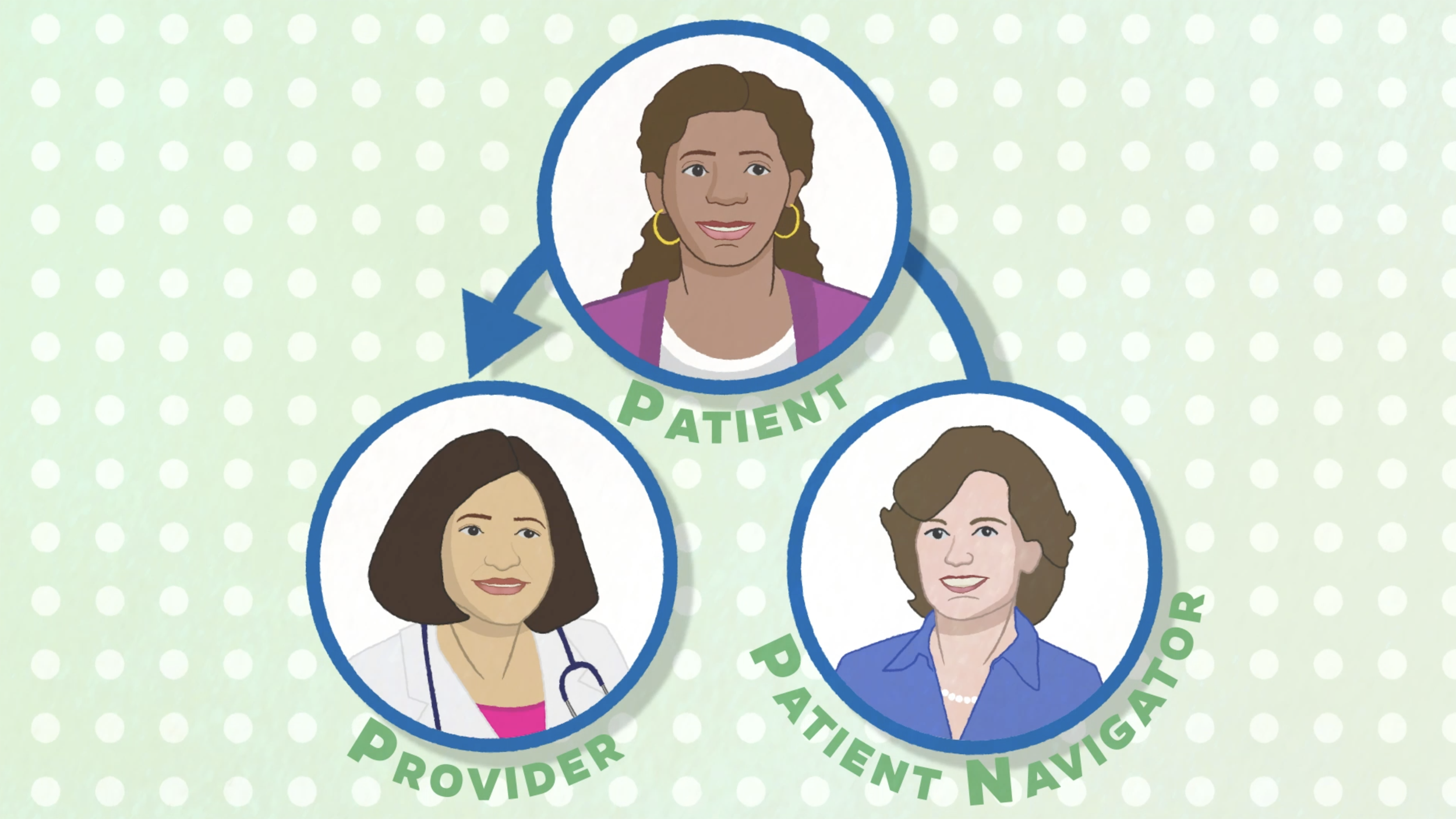
The Johns Hopkins Women’s Wellness and Healthy Aging Program is a new model for delivering midlife women’s healthcare that offers streamlined scheduling, interdisciplinary coordination and communication, a patient navigator to help establish patient’s relationships with providers in multiple specialties, and a personalized patient roadmap for healthy aging.
The rise in mobile communications over the last decade has revolutionized the way we access information. Social media has become an important platform for reaching patients, particularly underserved populations (Welch et al. 2016). Attention spans are shorter, making brief explainer videos an effective way to educate a patient population (Krämer and Böhrs 2016).
The research question this project addresses is whether brief explainer videos and illustrations on social media are effective at educating patients about this new model of integrated care. Patient stakeholder feedback was solicited at the beginning of the project to determine the most burdensome aspects of accessing quality healthcare and the health topics of most concern to middle-aged women. Subsequent rounds of stakeholder feedback were integral to the iterative process of creating the visuals and determining their efficacy. Two animations and a suite of 20 illustrations were created using the Adobe Creative Suite addressing health concerns of the perimenopausal population such as healthy aging, reducing risk of cardiovascular disease and cancer, maintaining mental health and cognitive function, and treatment of menopausal symptoms.
Patient stakeholder feedback determined that short explainer videos and illustrations on social media can be an effective tool to educate patients. Educated and engaged patients often have better health outcomes (Paterick et al. 2017). This new model of healthcare delivery and mode of educating patients have the potential to improve healthcare outcomes and decrease healthcare costs for women during their post-reproductive years.
Thesis Student
Preceptor
Department Advisor
Teaching MRI Physics: Creating media with a focus on the Radiology CORE Exam :: EMILY WU
MRI physics is an important component of radiology training and the American Board of Radiology CORE exam. There is a need for improved learning resources on the topic, but current literature does not provide much information about what types of study materials residents value or creating effective media for teaching MRI physics. This thesis project explores the MRI physics resource needs of radiology residents, creates new media based on the results, and establishes the basis for a future study to test its efficacy.
An online needs assessment survey was created and distributed to current members and recent graduates of the Johns Hopkins diagnostic radiology residency program. Respondents reported that current MRI physics resources were confusing and lacked diagrams and animations. The results indicated that residents desire resources with appealing visuals and simplified details. Questions banks and practice questions were consistently rated as the most helpful resources, but webpages with animations, videos, and webpages with diagrams were also ranked highly.
We created a 10-minute animation introducing the fundamentals of MRI physics, incorporating feedback from the needs assessment. This project aims to fulfill the need for visually appealing MRI physics resources to help residents learn these concepts, and to inform educators and content creators about how to improve the quality of educational materials on the subject. After this project, we have also planned for a follow-up study to evaluate the thesis animation in comparison to other media modalities, with the goal of evaluating how radiology residents best prefer to study for the CORE exam.

Educating Patients: Communicating the Gut-Brain Connection in Parkinson’s Disease using Multimedia :: SUSIE YUN

Educating Patients: Communicating the Gut-Brain Connection in Parkinson’s Disease using Multimedia © 2021 Susie Yun
Parkinson’s disease (PD) is a type of synucleinopathy that is characterized by abnormal accumulation of α-synuclein (α-syn) aggregates in neurons (Challis et al. 2020). The misfolded α-syn triggers self-aggregation into neurotoxic amyloid fibrils that can be transmitted from cell to cell, ultimately causing neurodegeneration (Kim et al. 2019). Mounting evidence suggests that for a majority of PD patients, this process can originate in the gut and ascend to the brain via the vagus nerve (Kim et al. 2019). Once in the brain, α-syn pathology propagates to reach the midbrain leading to motor dysfunction.
Since PD is mostly known as a motor disorder, its non-motor symptoms often go undetected. By the time patients begin exhibiting motor symptoms, the disease is usually advanced to the extent that only symptoms can be treated (Dawson et al. 2019). Recent studies suggest that non-motor symptoms, particularly those associated with gastrointestinal (GI) dysfunction may appear as many as 20 years prior to neurological symptoms (Challis et al. 2020). It is important for the public, especially those with chronic GI complications to understand how prolonged GI symptoms may indicate early-stage PD.
However, two factors currently hinder education in this area: (i) scientific research papers are difficult to understand particularly for readers without a scientific background; and (ii) current visual resources for this topic are limited to schematic, often confusing diagrams and inaccurate anatomical and molecular images. In particular, spatial relationships between the gut and the brain are difficult to depict in 2D. And, no effective didactic 3D visualizations exist to address this important public health topic.
I propose an interactive platform of (i) a foundational 2D teaching module on the scientific background and pathogenesis of PD, and (ii) a narrative 3D animation highlighting recent studies of the gut-brain connection in PD. Learners will navigate at their own pace through the introductory learning module prior to viewing the more complex material.

Thesis Student
Department Advisor
2021 Thesis Presentations
The Thesis Presentations of the Class of 2021 was held on April 16, 2021, 3:00-5:00 PM (EDT). This year’s presentation were held remotely with a Live broadcast to the Department’s YouTube Page. The students presented to their faculty and preceptors.
![]()
Alumni, students, and the public were invited to participate through YouTube Live. Guests typed questions into the Chat window of the Live Broadcast, and a special Q&A was held at the end of all presentations to answer posted questions.
Links to the YouTube Live event will be shared to the Department’s social media accounts: Facebook, LinkedIn, Instagram, and Twitter.
2020
Class of 2020 – Thesis Research
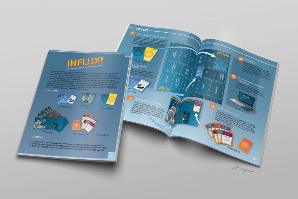
Creating a Game-Based Learning Tool for Optimizing Intra-Hospital Disaster Response :: NOELLE E. BURGESS
Preparing for disasters such as Mass Casualty Incidents (MCIs), whether of natural or human origin, are of increasing concern. When these incidents occur, hospitals are overrun with an influx of new patients and day-to-day operations are interrupted. Concurrently, increased demand for intensive care resources can outpace the ability of hospital staff to organize appropriately. In such events, efficient management of space (emergency rooms, operating rooms), medical staff, and supplies (blood, hospital beds) is imperative for reducing injury, illness and saving lives.

Game Board © 2020 Noelle Burgess
The Johns Hopkins Bloomberg School of Public Health recently identified key disaster categories for which United States hospitals need to be prepared: Large-scale natural disasters, complex MCIs, and catastrophic health events. Each category poses its own set of challenges for effective management of operations and resources.
During a complex MCI, demand for space, staff, and supplies quickly expand beyond the bounds of Emergency Departments (ED). Although many training tools exist for both the field and ED, surprisingly few exist for hospital-wide disaster response. Programs that do exist come in the form of infrequent, costly, and time-intensive large-scale simulations. A game-based instructional modality was developed to practice the medical knowledge, teamwork, and complex decision making required by multidisciplinary teams. As part of this research, a physical board game was developed using an iterative design approach. Additionally, a wireframe prototype of a complimentary digital version was developed using the Waterfall design approach. Both these options were chosen to reduce training costs, and increase training frequency and efficacy compared to large-scale MCI simulations.
The purpose of this project is to create an accessible and cost-effective game-based disaster readiness training tool specifically for intra-hospital personnel. The desired outcome is to increase efficiency and effectiveness of intra-hospital disaster response for MCI. For this project, a mass-shooting scenario has been selected as the focus of gameplay.
Rule Set © 2020 Noelle Burgess
Thesis Student
Department Advisor
Prostate Carcinogenesis: Depicting a Proposed Pathway for Prostate Cancer Precursor Lesion Development Using 3D Animation :: WILLIAM GUZMAN JR.
Prostate cancer is one of the most common malignant neoplasms among men in Western countries (De Marzo et al. 2017). In 2020, there will be an estimated 191,930 American men diagnosed with prostate cancer, an almost 10% increase from 2019 (Siegel et al. 2020). Visual resources for understanding of the pathogenesis of prostate cancer development, and specifically the development of prostate cancer precursor lesions, are limited. A visual void exists between prostate cancer Pathologists presenting histological findings and those with differing prostate cancer specialties. Furthermore, prostate cancer is typically studied using two-dimensional microscopy slides, and it is often difficult to explain new three-dimensional spatial hypotheses and findings to those outside of the pathology field.
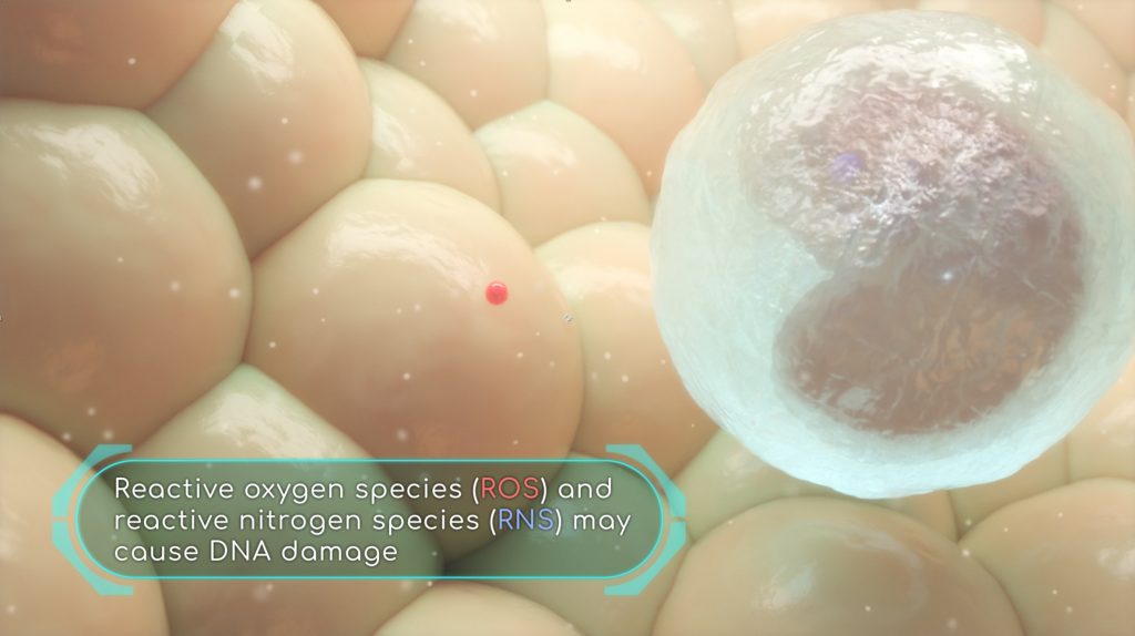
ROS and RNS may cause DNA damage © 2020 Willliam Guzman
Recent pathological evidence suggests a new role for prostate infections and inflammation in prostate cancer development. This novel hypothesis has the potential to challenge the dogma that high grade prostatic intraepithelial neoplasia (HGPIN) serves as the direct precursor to prostate cancer development. Rather, new molecular pathologic evidence indicates that an inflammation-associated lesion termed proliferative inflammatory atrophy (PIA) may directly transition to prostate cancer. 3D visualization of prostate cancer is novel and important in itself due to the multi-focal and atypical pattern of growth, and it is consequently challenging to communicate this new prostate cancer precursor lesion development model.
To address the lack of comprehensive histological research representation, a 3D mechanism of disease animation was constructed which portrays a new proposition for precursor lesion development using novel radical prostatectomy specimen data. The animation outlines and describes the harmful effects inflicted upon luminal epithelium caused by bacterial toxins, such as colibactin, as well as oxidants produced by immune cells induced by chronic inflammation. By improving visual understanding of histological data, this 3D animation provides a platform to further clarify the current knowledge of prostate cancer in the context of bacterial infections and chronic inflammation.
Thesis Student
Preceptors
Visualizing Cochlear Specializations that Enhance Protection of Hearing Function in Bats :: JAMIE LYNN PETERSON
The cochlea is a fluid-filled coil of the inner ear that transforms sound waves into electrical signals for processing in the brain. The mammalian auditory system is most commonly studied using the mouse, Mus musculus, due to anatomical similarities of the cochlea between auditory generalists, such as mice and humans. Auditory specialists, such as bats, exhibit unique resistance to age-related hearing loss, or presbycusis. This adaptation enables bats to navigate while flying with echolocation throughout their lifetime. Studying comparative cochlea anatomy can aid in understanding specializations of the mammalian auditory system and hearing loss among species. There is a significant gap in available educational resources for comparative cochlea anatomy focusing on bats and mice.
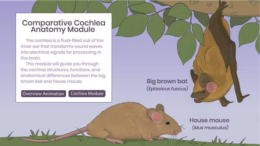
Comparative Cochlea Anatomy Module © 2020 Jamie Peterson
The purpose of this project was to develop an interactive educational resource for comparative cochlea anatomy of the big brown bat, Eptesicus fuscus and Mus musculus with a 3D overview animation depicting labeled cochlea models. Segmentations of histological and micro-CT data were modified and sculpted to build idealized anatomical models suitable for teaching purposes. A separate section of the interactive allows the user to explore comparative cochlear anatomy of bats and mice as related to hearing loss. The user interface and interactivity were coded to allow exploration of bat and mouse cochlea regions and intuitive navigation between sections about specific anatomical structures and bat hearing loss research. The results of this project provide a didactic and accessible visualization for auditory researchers, graduate students, and lay audiences to review basic cochlear anatomy, compare cochlear anatomy of bats and mice, and strengthen their understanding of human age-related and noise-induced hearing loss.
Comparative Cochlea Anatomy Module and 3D Models
Thesis Student
Preceptors
Department Advisor
3D Reconstruction of Fossilized Skull of South American Miocene Monkey Homunculus patagonicus: An Augmented Reality for Field Application :: KELLYN SANDERS
For most Miocene taxa, primate fossil evidence consists of broken cranial bones, teeth, and jaws. Studying these fossils is difficult due to the damage and distortion during geological stress. During fossilization the soft tissue preservation of these specimens is usually nonexistent. Homunculus patagonicus is an unusual primate from the Miocene epoch (~17 million years old) of extreme southern Argentina. The first associated cranium and mandible of this species will allow the most complete reconstruction of the adaptations of any early platyrrhine. To reconstruct the diet of such extinct mammals, the jaws, skull and muscles of mastication provide insight into how food properties influence skull morphology over evolutionary time (Perry, 2018). This project allows the use of comparative anatomy to learn how H. patagonicus lived, and its relationship to its environment through an examination of dietary adaptations.
Using extant analogs, correlations can be made between muscle and bone dimensions providing informed inferences about feeding behaviors in fossils. Inferences can be validated because, “Diet and mastication are closely tied to hard anatomy” (Perry, 2008). Thus, access to data from living analogs makes reconstructions of mastication especially justifiable for early primates (Perry, 2008). This data can be then used to recreate the magnitude and orientation of the force produced by the adductor muscles. These variables can be used to better understand the properties of foods and how they relate to food processing anatomy and behavior (Perry et al. 2011).
This project leverages digital visualization techniques to provide an interactive application using the digital fossil reconstruction of Homunculus patagonicus. The reconstruction of the skull and jaw adductor muscles are implemented through an interactive iOS application in addition to the original CT fossil data, extant distribution maps, and primate phylogeny. This application will not only provide researchers, students and the general public a learning resource but will also contribute to the fields of virtual paleontology, biocommunication and plastic surgery, especially facial reconstruction.
Thesis Student
Preceptors
Department Advisor
Visualizing HOPE: Encouraging HIV-Positive Organ Transplantation Using Novel Modular Animations :: MORGAN SUMMERLIN
HIV+ candidates in need of organ transplantation face an increased risk of mortality while on the organ waitlist and decreased access to transplantation compared to HIV-uninfected (HIV-) candidates. Fortunately, HIV+ donor organs can now legally be transplanted into HIV+ recipients, thanks to the HIV Organ Policy Equity (HOPE) Act of 2013, which reverses an outdated ban and unlocks a pool of organs from an estimated 300-500 HIV+ deceased donors in the US annually. However, challenges in the current organ donation system and stigma against HIV present barriers to HOPE implementation and the potential for life-saving HIV+ donor to HIV+ recipient (HIV-to-HIV) organ transplantation. In collaboration with transplant surgeons, infectious disease healthcare providers, organ donation community consultants, and medical illustrators, a novel animation workflow was developed to create HOPE education materials. The animations, targeted to potential donors and recipients with HIV, healthcare providers, and professionals in the organ donation community are intended to promote awareness and ultimately increase the rate of HIV-to-HIV transplantation.
To efficiently communicate to these critical audiences, the animation process was streamlined by combining overlapping information into reusable components and addressing each audience with a customized call-to-action clip. The results of this project provide two individual animations addressed to (1) people living with HIV and (2) professionals in the organ donation community. The animations consist of reusable clips that explain the background information of the HOPE Act, the potential biological risks of HIV-to-HIV transplantation, and a concluding statement that reminds the viewer that their participation is essential to continue the success of the HOPE Act. Each animation concludes with an individualized call-to-action segment that inspires viewers to make a change.
By informing these critical audiences through inclusive and educational animations, this project aims to reduce stigma in HIV+ organ donor referrals and registration and to encourage participation in HIV-to-HIV transplantation.
Thesis Student
Personalizing Hospital-based Stroke Education: Designing a Novel Recovery App to Prepare Stroke Patients for the Transition Home :: HELEN TANG
Stroke is a leading cause of adult disability in the United States. It is a complex disease for which timely intervention and treatment has the highest yield. Individualized patient education is crucial for optimizing recovery, preventing recurrence, and improving patient outcomes. Current hospital-based education is largely paper-based and suboptimal for meeting the varied, yet precise educational needs of the stroke patients and their care partners (carers). To augment the current education program, the multidisciplinary stroke team at the Johns Hopkins Comprehensive Stroke Center is building a mobile app that will provide individualized, interactive, and accessible education to prepare hospitalized stroke patients and their carers for the transition from hospital to home. This thesis project lays groundwork for the development of the app by designing its overall structure and navigation, and by prototyping specific paths demonstrating key features and functions of the app.
User-centered methodology was implemented to focus each stage of app design on the fulfillment of unmet learning needs of stroke patients and carers in acute hospital care and after hospital discharge. Key identified needs were synthesized into four enabling objectives and the underlying information architecture that informed the scope and structure of the app. Digital prototypes of seven key tasks that translated the abstract groundwork into concrete visuals were developed in an iterative and collaborative process with stakeholders.
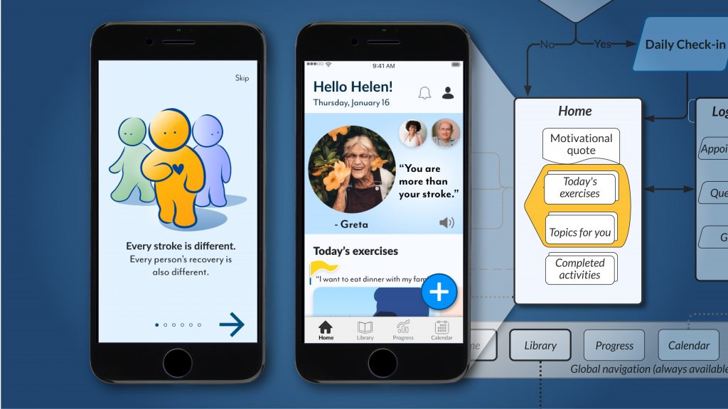
Designing a Novel Recovery App to Prepare Stroke Patients for the Transition Home © 2020 Helen Tang
The core features designed were (i) personalization of daily educational content, (ii) actionable recovery goal-setting, (iii) progress tracking, and (iv) improved two-way communication between patient and care team. Corresponding information architecture, interactive digital prototypes, and a model for progressive personalization were constructed. Together, these contributions provide the foundation for development of the first iteration of the app, and serve as valuable communication tools for continued collaboration and planning between stakeholders. The user-centered methodology imparted structure and strategy to the design process, while iteration enabled adaptability to new insights. Frequent usability testing, inquiry, and collaboration with stakeholders were essential to design refinement. The continued use of these methods during app development will maximize usability and efficacy of this novel personalized educational resource for early stroke recovery.
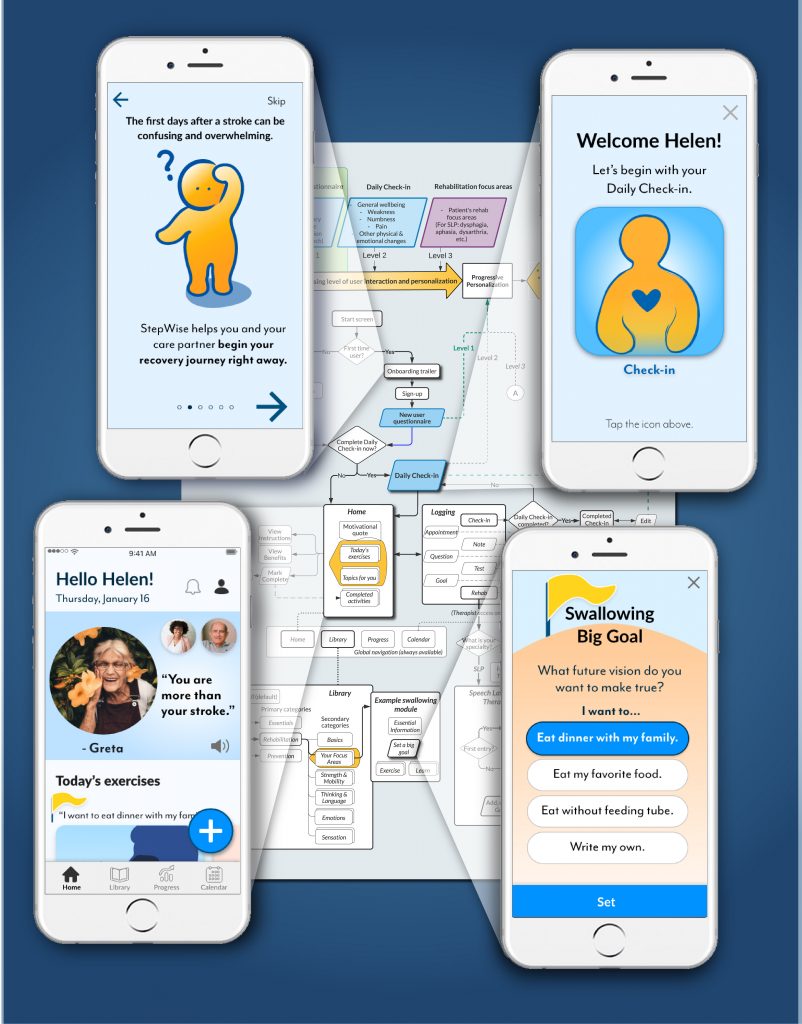
Personalizing Hospital-based Stroke Education © 2020 Helen Tang
Thesis Student
Department Advisor
Optimizing e-Learning in genetics: creating and comparing three categories of multimedia :: JENNY WANG
Online learning is rapidly expanding in the United States. One feature of online learning is the increased use of animations, especially in the sciences. However, there are contradictions within the literature regarding the effectiveness of animations in scientific education. Some studies claim that animation is the best modality for teaching scientific topics, while others have shown that it increases cognitive load, leading to reduced effectiveness. This thesis will test these opposing positions by measuring the effectiveness (retention and engagement) across three types of multimedia that we created: (i) a 6 minute 38 second traditional 2D animation, (ii) a 6 minute 43 second whiteboard animation, and (iii) an 8 minute 11 second PowerPoint video edited together from lecture videos. This three-way comparative approach will determine intrinsic differences and similarities across multimedia.
We recruited study participants from Amazon Mechanical Turk (N=168), split into six groups of 28 differentiated by video order. Retention and engagement scores were collected via survey in JHM Qualtrics. Using single factor ANOVA, we found no difference (p < 0.05) among the three modalities for retention. However, whiteboard animation performed better with word recall than the other two videos, suggesting that simultaneous narration with written text leads to better learner outcomes. We also found that the two animation formats performed better (p < 0.05) than the PowerPoint lecture for engagement (enjoyment, attention, understanding). This project aims to provide insight for e-Learning creators into which modalities work best for engaging and teaching learners while also considering monetary costs.
Thesis Student
Department Advisor
2020 Thesis Presentations
The Thesis Presentations of the Class of 2020 were held on April 17, 2020, 3:00-5:00 PM (EDT). This year’s presentation were held remotely with a Live broadcast to the Department’s YouTube Page. The students presented to their faculty and invited alumni, students, and staff from Johns Hopkins Medicine as well as the public to participate through YouTube Live. Links to the YouTube Live event were be shared to the Department’s other social media accounts: Facebook, LinkedIn, Instagram, and Twitter.
Order of Presenters: Jamie Peterson, Helen Tang, Jenny R. Wang, William Guzman Jr., Kellyn Sanders, Morgan Summerlin, and Noelle Burgess.
2019
Class of 2019 – Thesis Research
Teaching the Cellular and Molecular Basis of Breast Cancer Metastasis: A Novel Workflow for Incorporating Time-lapse Microscopy Data into 3D Animation :: BRITTANY BENNETT
Time-lapse confocal microscopy and organotypic 3D culture allow biologists to capture 3D movies of cells moving in real time. The Ewald Lab at Johns Hopkins School of Medicine developed this method to determine how molecular variables affect the growth of breast cancer tumors and cells’ ability to metastasize. The results support a new model of breast cancer metastasis, called Collective Epithelial Metastasis (CEM). Existing visuals of CEM are limited to microscopy and schematic model figures. Although informative to biologists, these are not intuitive to non-specialist audiences such as patient advocates and research investors. There is a need for visuals that explain the molecular and cellular basis of CEM within an anatomical context, and make conclusions of complex research more accessible.
The animation teaches the role of two proteins, E-cadherin and Keratin-14, during collective invasion and dissemination. The visual challenge was to contextualize molecular concepts for an audience that first needs introduction to mammary gland anatomy, histology, and epithelial cancer definitions. Learning objectives, a script, and twenty-four page partial-color storyboard were created to teach these concepts in an appropriate level of detail. A website was developed to display the animation and provide additional information and citations.
The technical project goal was to incorporate the Ewald Lab’s time-lapse microscopy datasets into an educational animation. Selected datasets show cellular events that correspond to storyboarded scenes. Volumetric data was converted into 3D surface meshes in Bitplane Imaris and imported into MAXON Cinema 4D with an OBJ Sequence Importer plugin. In Cinema 4D, the “re-animated” surface meshes were modified before being merged into a scene of the mammary duct microenvironment. Other data-derived 3D models created for the animation include: 1) breast anatomy and a metastatic tumor, segmented from DICOM data and 2) a ductal tree created in Cinema 4D, based on mouse mammary tissue.
This project demonstrates that 3D time-lapse microscopy datasets can be incorporated into Cinema 4D and blend within the built anatomical and histological scene. The merging of data-derived animation with artist-created animation can improve scientific communication to audiences outside of cell biology.

Thesis Student
Preceptor
Department Advisor
Morphology of Memory: Creating a web-based 3D interactive resource to teach the anatomy of the human hippocampus :: ALISA BRANDT

Morphology of Memory Interactive © Alisa Brandt, 2019
The hippocampus is a critical region of the brain involved in memory and learning. It has been widely researched in animals and humans due to its role in consolidating new experiences into long-term declarative memories and its vulnerability in neurodegenerative diseases. The hippocampus is a complex, curved structure containing many interconnected regions that consist of distinct cell types. Despite the importance of understanding the normal state of hippocampal anatomy for studying its functions and the disease processes that affect it, didactic educational resources are severely limited. The literature on the hippocampus is expansive and detailed, but a communication gap exists between researchers presenting hippocampal data and those seeking to improve their understanding of this part of the brain. The hippocampus is typically viewed in a two-dimensional fashion; students and scientists have difficulty visualizing its three-dimensional anatomy and its structural relationships in space.
To improve understanding of the hippocampus, an interactive, web-based educational resource was created containing a pre-rendered 3D animation and manipulable 3D models of hippocampal regions. Segmentations of magnetic resonance imaging data were modified and sculpted to build idealized anatomical models suitable for teaching purposes. These models were animated in combination with illustrations and narration to introduce the viewer to the subject, and the completed animation was uploaded online and embedded into the interactive. A separate section of the interactive allows the user to rotate the models, hide and show different regions, and access additional explanatory text. The user interface and interactivity were coded to allow exploration of hippocampal regions and navigation between sections of the resource. The results of this project provide a didactic and accessible visualization for graduate students, researchers, clinicians, and other individuals involved in neuroscience. The animation and interactive models allow users to reinforce their understanding of 3D hippocampal anatomy and connectivity. By improving visual understanding of the hippocampus, this project aims to advance the communication and scientific study of hippocampus-related topics, such as epilepsy and Alzheimer’s disease.
Morphology of Memory
Thesis Student
Preceptor
Department Advisor
Autopsy Brain Removal Training Using Virtual Reality Simulation :: INSIL CHOI
Hospital autopsy is the only method of confirming diagnoses for neurodegenerative disease such as Alzheimer’s and Lewy body disease, despite advanced diagnostic technologies. However, the number of hospital autopsies has steadily declined, due to changes in hospital accreditation requirements, lack of reimbursement, and other factors. Consequently, it is challenging to train autopsy assistants, pathology residents and neuropathology fellows to become competent in evisceration and dissection techniques, as there are few opportunities to observe and perform hospital autopsies. The procedure for autopsy brain removal is particularly challenging because: (i) incorrect cutting planes and depth of cut during opening of the cranium can lead to inadequate exposure of, or damage to, brain tissue, and (ii) there may be initial hesitation to perform the procedure due to proximity to the face. These challenges are compounded because current teaching resources are limited to drawings (which lack key spatial and volumetric cues) and photographs (which provide only a single view, and may have superfluous information); no physically practice of the procedure is involved.

Autopsy Brain Removal Training © Insil Choi, 2019
To address this deficit, a virtual reality (VR) simulation application was developed to teach proper methodology in performing steps of the autopsy brain removal, and as a proof-of-concept for a complete hospital autopsy VR simulation. The simulation provides an immersive VR interactive experience within an Oculus Rift platform. It uses realistic virtual patient models created from surface 3D scans of a real person, data-driven 3D models of anatomy, sound effects, and haptic responses within a VR autopsy suite. The simulation also features real-time visual feedback and evaluation of user performance, to assist improvement of skills and knowledge during the step of opening the cranium with an oscillating saw.
This thesis documents the process of developing the VR simulation, in particular the creation and use of i) 3D models of patient, cranium and brain, ii) interactions between instruments and patient in VR, iii) in-game feedback: visual, sound and haptic, and iv) user interface (UI) interaction in VR.
Autopsy Brain Removal Training – Virtual Reality Simulation
CAUTION: The above video contains images and sounds of a simulated autopsy that may be disturbing to some viewers.
Thesis Student
Preceptor
Department Advisor
Mapping the Mechanobiome: Novel mathematically-derived 3D visualization of the cellular mechanoresponsive system for interactive publication :: CECILIA JOHNSON
Mechanical forces, ubiquitous in biological settings, are major determinants of cell fate; they should not be considered a detail applicable to specialized circumstances but rather a vital component of cell biology. To sense, respond, and generate both intracellular and extracellular mechanical forces, cells contain a highly integrated and dynamic network of macromolecules throughout the cell. The Robinson Lab at the Johns Hopkins School of Medicine developed the term “mechanobiome,” to describe and categorize that network of macromolecules. At the interface of cell biology, physics, and engineering, the concept of the mechanobiome provides researchers a systems-level understanding of the extensive contributions of physical force and mechanical cell properties on cell morphology, differentiation, physiology, and disease. Although numerous diseases, including cancer, cardiovascular disease, and chronic obstructive pulmonary disease, develop from abnormal cell mechanics, the mechanobiome is rarely explored as a novel source of therapeutic targets. Increased understanding of the mechanobiome will enhance understanding of normal biological machinery and ultimately lead to new pathways for targeting disease.
To address the lack of comprehensive, accurate visualizations of the mechanobiome, two novel theoretical 3D models of the mechanobiome were developed: one at the cellular level and one at the nanoscale level. By integrating published data on components of the mechanobiome, such as crystal structures, macromolecule concentrations, and polymer dissociation constants, a proportionately accurate visualization of the cell’s mechanical system was produced. A platform was prototyped to present these novel 3D visuals as interactives on an accessible web-based educational resource, “Mapping the Mechanobiome.” The resource also provides review-style descriptions of fundamental concepts in mechanobiome research with accompanying visual media generated from the mathematically-derived models.
This resource will contribute to discussions on the forefront of mechanobiome research, provide a comprehensive understanding for new researchers in the field, and advance research efforts by highlighting the significance of fundamental mechanical properties. The novel, mathematically-derived models have the potential to reveal aspects of the mechanobiome not previously considered due to the lack of accurate visualization of the full working system. This resource provides a platform to further enhance our understanding of the role of mechanics in health and disease.


Thesis Student
Preceptor
Department Advisor
Creating a Character Animation-based Interactive Frailty Model to Support Better Primary Care Implementation and Planning for Older Adults :: LOHITHA KETHU
As global populations age, it is imperative for clinicians and researchers to understand and apply measures of frailty syndrome to support healthy aging. Frailty syndrome is defined as a clinically recognizable state of increased vulnerability resulting from aging-associated declines in reserve and function across multiple physiologic systems, compromising the ability to cope with every day or acute stressors. Frailty predicts surgery outcomes, waitlist times, cancer therapy tolerance, disability, institutionalization, morbidity, and mortality. Insufficient understanding of the complex nature of frailty from molecular changes, to physiology to clinical changes, is a major challenge for clinicians and researchers that focus on uni-dimensional (one factor at a time) molecular pathways to late-life decline connections. In a collaboration between clinicians, frailty researchers, and medical illustrators, a multidimensional, character animation-based resource was built, which articulates the aging and frailty-related changes from the cellular level to the clinical level, and provides visualizations that facilitate an understanding of a multidimensional, holistic frailty theory. With guidance from the Frailty and Multisystem Dysregulation Working Group at the Center on Aging and Health, a two-part animated, web-based, interactive 2D and 3D module was created to convey both “2D” clinical theory and “3D” real patient experience, created in ZBrush and Cinema 4D. The interactive portion of the module was created through Blender and Verge3D.

Thesis Student
Preceptor
Department Advisor
Tablet-Based Training for Open Reduction Internal Fixation: A Model for Complex Pelvic Trauma Surgery :: VONDEL MAHON
Open Reduction Internal Fixation (ORIF) surgery of the pelvis is a highly complex procedure that should only be performed by the most highly skilled and experienced orthopaedic surgeons. To develop the appropriate skills, surgeons must complete at least five years of orthopaedic residency training including mentorship and exposure to a wide array of surgical cases. The expectation of residency training is that new surgeons will develop the skills and competencies, and attain the experience that will enable them to be proficient in their future role. However, the current training system is outdated and often does not provide sufficient opportunities to master skills, particularly those that require surgeons to convert 2D information into 3D mental models. As a result, some orthopaedic surgeons may be inadequately prepared to perform ORIF surgeries.
This thesis explores the design of a tablet-based 3D interactive surgical training tool that teaches ORIF procedures. It will allow orthopaedic residents to improve geometric and spatial understanding of fractures, skeletal anatomy, and surgical hardware, in the context of ORIF surgery; enable investigation of optimal positioning of plates and screws, and allows users to confirm their placement by means of x-ray views; promote higher levels of confidence in reduction and fixation of unstable pelvic fractures. The overarching goal of this interactive surgical training tool is to enable surgical residents to practice ORIF surgery on virtual patients created from Digital Imaging and Communications in Medicine (DICOM) derived anatomical models and virtual surgical hardware. This approach allows DICOM data to be developed into virtual cases and used as training material, thus allowing residents to increase their exposure to relevant surgeries and improve their skills in ORIF procedures.
The research project is a work in progress. It is part of a larger study at Johns Hopkins University School of Medicine. The interactive surgical training tool has the potential to truly impact the quality of patient care by helping orthopaedic residents gain the skills and experience necessary while using simulated cases. Once fully tested and developed, this interactive surgical training tool has the potential to be used by orthopaedic residency programs around the globe.

Thesis Student
Preceptor
Department Advisor
2019 Thesis Presentations
The Thesis Presentations of the Class of 2019 was held on April 19, 2019, 3:00-5:00 PM, in the Chevy Chase Auditorium of the Johns Hopkins Hospital. The students presented to an audience of faculty, alumni, and students from Johns Hopkins Medicine, and the public. The recording was broadcast through Facebook Live.
Order of Presenters: Brittany Bennett, Alisa Brandt, Lohitha Kethu, Insil Choi, Cecelia Johnson, and Vondel Mahon.
2018
Class of 2018 – Thesis Research
Creating an Interactive Algorithm for Improved Management of Difficult Airways in Micrognathic and Retrognathic Infants :: LAUREN RAKES
Newborns with conditions such as Pierre Robin sequence, Treacher Collins syndrome, and Goldenhar syndrome are born with underdeveloped mandibles, a condition known as “micrognathia”. Micrognathic and retrognathic (posterior positioned jaw) patients face challenges and risk of injury when anesthesia is required, due to a hypoplastic mandible, posterior displacement of the tongue, and other facial anomalies that ultimately cause airway obstruction and difficult intubation.

Creating an Interactive Algorithm for Improved Management of Difficult Airways in Micrognathic and Retrognathic Infants © Lauren Rakes, 2018
Currently, a “Difficult Airway Algorithm” is used to guide physicians to make the best choices during adult intubation, depending on the particular challenges and dynamics of the case. However, there are no widely available or accepted difficult airway algorithms for micrognathic infants. Furthermore, simulation models exist for practicing intubation on newborns, but only one model is available with micrognathia and is not readily available at many medical teaching facilities. Lack of training resources and contact with patients with this condition prevent physicians from acquiring the experience necessary to adequately care for these infants.
An interactive web application was created to provide an accessible way to increase exposure to the anatomy of micrognathic newborns and the recommended techniques for intubation. A unique interactive algorithm within the application was developed specifically for the treatment and airway management of children with micrognathia. Interactivity allows the user to explore different paths within the algorithm and learn additional information through text, illustration, and animation. Additionally, a separate section of the application allows for exploration and deconstruction of a 3D model of a micrognathic patient, allowing for reinforcement of anatomical and spatial concepts. Together the unique interactive flowchart, the 3D anatomy viewer, and an additional background information section with supplemental text and illustrations, provide a useful overview for users that anticipate the need to manage a micrognathic airway. This project will contribute to the safety of these children inside and outside the operating room by increasing clinician’s exposure to the different options available during intubation. The workflow and foundation of this project has the potential to be expanded and applied to a variety of medical scenarios.
Improved Management of Difficult Airways
Thesis Student
Department Advisor
A novel web-based public outreach tool for promoting the critical role of fundamental research in human health: Incorporating scrolling triggered video playback into the user experience :: TIANXING SHI
Fundamental research has played a significant role in human health and its impact continues to grow. As researchers probe deeper into cellular and molecular mechanisms of complex diseases like cancers, fundamental research has evolved into highly specialized subfields. These developments in research, however, have moved knowledge and understanding even further from the grasp of the general public. Meanwhile, not enough is done to effectively explain to the public why scientists do what they do. The subsequent communication breakdown between the science community and the public may cause decreased research funding, lack of interest in pursuing careers in science, and a growing ignorance on the part of the public regarding the value of fundamental science. This thesis seeks to bridge this communication gap by developing a public outreach program to clearly explain the principles of fundamental research and address its importance in improving human health.

Image – Critical Role of Fundamental Research in Human Health © Tianxing Shi, 2018
This thesis explores the effectiveness of making a public outreach program engaging and effective by employing a combination of visual representations, including 3D models, animations, 2D motion graphics, user interface and experience designs. The final product will be presented on an interactive video website using a recently developed JavaScript coded technique called “Scrolling Triggered Video Playback”. This technique allows viewers to play and playback the videos by scrolling up and down based on their reading pace. Content of the word story and art assets is based on research projects carried out in the lab of Douglas Robinson in the department of Cell Biology at Johns Hopkins University School of Medicine. The Robinson Lab studies cell mechanics, including mechanoresponsive behavior of cells, and applies the concepts they observe to developing novel therapeutics for complex diseases, including cancer and chronic obstructive pulmonary diseases (COPD).
Expected benefit of this public outreach program is to effectively communicate the value and importance of fundamental research to the public. The goal of the program is to assist viewers in understanding the general concepts behind fundamental research by using current topics from the Robinson lab as examples, and in doing so cultivate an appreciation of the relevance of science to people’s lives.
Promoting the Critical Role of Fundamental Research
Thesis Student
Preceptor
Department Advisor
Resurrecting an Ancient Bite: Virtual Model Sheds Light on one of the Earliest Primates :: AMANDA SLADE
The first true primates in the fossil record are known from near the Paleocene-Eocene boundary, approximately 55 million years ago. The prevailing evidence suggests that these primates diversified rapidly, making the sequence of events which make up their evolutionary history challenging to navigate. (Kay et al., 2004; Rose, 2006). A powerful tool for enhancing the understanding of primate origin is deciphering early primate chewing anatomy, as it can help paleontologists to infer a range of features including diet, body form and function and evolution. (Perry, 2008). The study of such ancient anatomy is inherently difficult, however, due to their fossil remains often being damaged and distorted, having undergone eons of geological stress. Furthermore, soft tissue is usually completely absent from the fossil remains of this time period.

Resurrecting an Ancient Bite © Amanda Slade, 2018
Perry and colleagues (2015; 2018), however, have developed a rigorous muscular reconstructive techniques using mathematical estimates of muscle size based on muscle-bone correlation in living analogs. These techniques can radically enhance the reconstruction of a specimen but can be hindered by sediment obscuring the necessary osteological measurement points. Additionally, these estimates, although novel, are entirely static numerical reconstructions making their implications and plausibility difficult to visualize.
This project used virtual visualization techniques to facilitate both the accessibility and dynamic reconstruction of chewing muscle data acquired from a specimen of one of the earliest primate species, Smilodectes gracils. The rigorous restoration of the virtual skull allowed for novel jaw adductor volume data to be collected and subsequently visualized through an interactive web application, featuring the virtually reconstructed 3D skull, chewing musculature, and an animated chewing simulation which brings static numerical data to life. This project contributes to the fields of virtual paleontology and biocommunication by using visualization to both extract and dynamically display hard data. These tools helps demystify a portion the often convoluted and controversial discussion of primate origins, and have implications for understanding the ecological history of our own species.
Resurrecting an Ancient Bite
Thesis Student
Preceptor
Department Advisor
Stem Cell Orchestra: Interactive Didactic Animation for Cardiac Tissue Engineering :: SHAWNA SNYDER
A potential treatment of heart failure from myocardial infarction is to replace damaged tissue with a cardiac patch, a bioengineered construct of cardiomyocytes and stem cell-supported capillaries grown on a bioscaffold. Tissue engineers must have a thorough understanding of the cues that guide multiple cell types to form functional tissue. As the field of cardiac tissue regeneration develops novel protocols for stem cell-based therapies, visualizations that didactically convey in vitro spatiotemporal cell-cell interactions become increasingly important to provide to students and the general scientific community.

Cardiac Patch Cellular Microenvironment © Shawna Snyder, 2018
An interactive web-based animation and educational module, Stem Cell Orchestra, was designed and developed to introduce students to case-based examples of biomedical research that are directly related to fundamental tissue engineering principles. A conceptual flowchart was created to establish the navigational structure of the module and connections between educational topics. The module focused on visualizing the development of a cardiac patch as a model bioengineered tissue through the creation of a 3D animation depicting cell-cell interactions within a cardiac patch. Animation content was derived from confocal microscopy and transmitted light microscopy datasets in combination with the results of a literature review of current cardiac regenerative medicine techniques. The educational module presents the 3D animation within the context of supplemental educational material on mechanotransduction cues related to cardiac patch development.
This interactive animation platform introduces students to examples of primary research and accurately showcases tri-culture of a cardiac patch from cardiomyocyte elongation and synchronization to the formation of endothelial capillaries supported by the novel human adipose derived stem cells. Supplemental material contextualizes mechanotransduction presented within the animation. This project resulted in the development of a novel workflow and educational module that can be expanded to include additional tissue engineering concepts.
Stem Cell Orchestra: Building a Cardiac Patch
Thesis Student
Preceptor
Department Advisor
Communicating the Iterative Spiral Model of Healthcare Innovation
Using Narrative Animation :: TZIPORAH THOMPSON
In an industry dominated by established corporations, current trends show increased innovative technology for healthcare solutions coming from small start-up groups. These start-ups often germinate in an academic setting, relying on the guidance and resources of their educational institution. However, traditional engineering or clinical programs often do not address the requisite bridging between technological innovation and market application. This can lead to a potentially effective healthcare solution failing to reach its patients or users.

Iterative Spiral Model of Healthcare Innovation © Tziporah Thompson, 2018
In order to address this educational gap, the Johns Hopkins Center for Bioengineering, Innovation, and Design (CBID) developed a model for efficiently navigating the process of healthcare innovation. This model focuses heavily on iteration, stakeholder feedback, and business model de-risking. A narrative animation was created in order to effectively communicate this educational model. The animation introduces the application of the iterative model using pressure ulcers as an example clinical problem. Characters and storytelling were used to stimulate viewer engagement and concept retention.
The goal of this animation was to give potential innovators a starting point and mental model with which to further their understanding of the healthcare innovation process. Feedback from stakeholders was regularly incorporated throughout the animation development process in order to emphasize the
clarity and efficacy of concept delivery using narrative animation.
Future directions include providing first-time innovators with a comprehensive educational platform by consolidating important resources into a single webpage. This would include the finalized animation, along with an interactive module, and relevant case studies for reference.
Intro to Healthcare Innovation
Thesis Student
Preceptor
Department Advisor
Gender-Affirming Surgery: A Visual Resource for Patients, Relatives, and Healthcare Providers :: HILLARY WILSON
Gender dysphoria occurs when an individual’s psychological identity as a male or female conflicts with their biological sexual characteristics, resulting in anxiety, depression, and distress. Transgender individuals often face harassment, discrimination, and stigmatization. Discrimination and transphobic experiences in clinical settings can erode trust in the healthcare system by the transgender community, and by extension, negatively affect the health of the transgender patient.

Gender-Affirming Surgery: A Visual Resource © Hillary Wilson, 2018
Gender-affirming surgeries are being performed with increasing frequency. In 2014, the Affordable Care Act banned discrimination based on gender identity, and Medicare coverage was extended to include transgender surgical treatment. The number of transgender individuals seeking gender-affirming surgery is expected to rise.
Gender-affirming bottom and facial surgeries are complex, difficult to conceptualize procedures and require the patient to make decisions about permanently changing two intimate parts of the body: the pelvic anatomy and face. Failure of the patient to understand resulting anatomical changes and the implications those changes have for post-operative function and upkeep can lead to low post-operative satisfaction and patient outcomes. Patient-doctor communication is crucial for effective decision making by the patient. It is also important to educate potential downstream healthcare providers who may be unfamiliar with these procedures and the new anatomy of post-surgical patients.
Existing visual resources on gender-affirming surgery are limited; they often lack clarity, are inconsistent, or are not designed for patient education. Videos and photographs may be graphic and impractical for teaching purposes. A full color, gender and racially sensitive patient education resource was created with input from professionals at the Johns Hopkins Center for Transgender Health. It includes nine male to female vaginoplasty images, six female to male phalloplasty images, and four donor site images. Several images employ an innovative color-coding system to describe tissue repositioning. A ten-image facial feminization surgery interactive module was designed to educate patients on realistic post-surgical outcomes of six procedures that modify facial structure. The images, rich in detail and aesthetically pleasing, will facilitate doctor-patient communication, aid patient decision-making, and improve health outcomes and trust in the healthcare system for the transgender community.


Thesis Student
Preceptor
Department Advisor
2018 Thesis Presentations
The Thesis Presentations of the Class of 2018 were held on April 20, 2018, 3:00-5:00 PM, in the Chevy Chase Auditorium of the Johns Hopkins Hospital. The students presented to an audience of faculty and students from the Hopkins and Augusta University medical illustration programs, as well as alumni from Hopkins, other Hopkins faculty, and the public. The recording was broadcast through Facebook Live.
Order of Presenters: Lauren Rakes, Tziporah Thompson, Mary Tianxing Shi, Hillary Wilson, Amanda Slade, and Shawna Snyder.
2017
Class of 2017 – Thesis Research
A Patient-Friendly Web Resource for Vascular Anomalies: Establishing a Connection between Patients and Clinicians :: ELEANOR BAILEY
Patient education is fundamental to patient-centered care which emphasizes shared decision-making between patients and clinicians. Patients and families must have a sufficient understanding of health information to participate in health decision-making, and this understanding is garnered from accurate, reliable, and readable health resources accompanied by helpful visuals. With 84% of Americans accessing health information online, the importance of ensuring these resources are comprehended by consumers must be emphasized. If health care providers wish to communicate with the general public, they must ensure health information is written at a level understandable to the average American.
A 2016 study of online resources about vascular anomalies, including those from Boston Children’s Hospital, Cincinnati Children’s Hospital, and Johns Hopkins Hospital, revealed none of these resources are readable by the average American. The language and sentence structure are complex, commensurate with the reading level of high school or college graduates, while the average American reads at the seventh grade level. Additionally, few visual aids about vascular anomalies exist, and the primary visual representations shown are graphic patient photographs.
The lack of understandable resources about vascular anomalies hinders a shared decision-making process between patients or families and clinicians. By utilizing a combination of readability tests as well as the first standardized index of quality health information, known as DISCERN, a comprehensive and readable source of health information about vascular anomalies was developed. The diagnostic and treatment information was gathered from several experts in the field and vetted by the specialists at Johns Hopkins Hospital. This web resource contains text that is understandable to the average American and illustrations that show the condition without frightening patients.
A disparity exists between how patients and clinicians approach health information; this is apparent in how web-based patient resources are currently presented. To establish a connection between patients and clinicians through web-based patient education, the principles of design governing traditional visual communication must be applied in the virtual world and amended to include accessibility, readability, and organization in virtual space.

Thesis Student
Department Advisor
Designing Customized 3D Printed Models for Surgical Planning in Repair of Congenital Heart Defects :: SARAH CHEN
Congenital heart defects (CHD) present with a wide variety of structural anomalies that range in severity, creating a need for personalized, precision treatment. Recent advances in diagnostic imaging and 3D printing technology have enabled the creation of patient-specific models, which have revolutionized the understanding and management of these conditions.
The objective of this research was to explore each step of generating such prints, and to establish a novel workflow for creating CHD models. The research focused specifically on creating aortic arch models optimized for surgical planning for patients with hypoplastic left heart syndrome (HLHS), a CHD in which the left heart and aorta are severely underdeveloped. Like most CHDs, there is substantial variability in HLHS anatomy, and the 3D shape of the aortic arch after reconstruction is critical in determining proper cardiac output, justifying a need to create custom 3D prints for improved surgical outcome.
While several software options exist for segmentation, this project concentrated on evaluating the following six to segment anatomy: Mimics, inPrint, OsiriX MD, Vitrea, D2P, and 3D Slicer. Meshmixer and 3-matic were used to manipulate the data exported from the segmentation software, to create life-sized models of pre-operative anatomy, an approximation of desired post-operative anatomy, and a customized homograft patch for aortic arch reconstruction. The models were printed on a Stratasys Connex3 Object260 printer using Tango Plus flexible material to allow surgical suturing.
Although some models were segmented from CT acquired data, emphasis was placed on establishing methods for utilizing 3D ultrasound derived data. This alternative provides a safe, cost-effective, and accessible imaging modality without harmful radiation, contrast, or anesthesia in vulnerable pediatric patients. Additional proof of concept models were derived from 3D fetal cardiac ultrasound data, since the first stage operation for HLHS—as well as other complex CHDs—must often be performed days after birth. Customized 3D printed models have the potential to improve treatment planning, reduce procedure time and improve patient outcomes. Workflows proposed in this project facilitate a safer and more effective method for producing 3D printed models of a pediatric heart.

Thesis Student
Preceptor
Department Advisor
Virtual Reality Guide to Hospital Autopsy: Block Dissection of the Renal System :: DANIEL HERMANSEN
Hospital autopsies are a highly valuable source of pathological evidence for understanding the progression and nature of diseases. Autopsies can also serve as a healthcare audit, assuring that premortem diagnoses are complete and accurate. However, requests for hospital autopsies have continually declined since 1970. This has resulted in a lack of understanding of the benefits of autopsies among patients and clinicians, and a dearth of professionals trained to competently perform the procedure. With limited access to procedures and professionals qualified to train others, certification has become increasingly difficult. In addition, the training materials currently available are limited and do not allow one to physically practice the techniques involved in the autopsy procedure.
This study documents the methods used in developing the Virtual Reality Guide to Hospital Autopsy: Block Dissection of the Renal System. This virtual simulation serves as a training tool to teach proper post-mortem examination technique through immersive user interaction. The simulation covered in this study will serve as the foundation and proof-of-concept for a future expanded program covering each of the five anatomical block dissections involved in a standard autopsy. This study will address the creation of one of those blocks, the renal system, in particular the creation and utilization of 1) a virtual user interface, 2) interactive virtual instruments, 3) accurate virtual models of renal system anatomy, 4) realistic tissue reaction of organic models with instrument interaction, and 5) a demonstration of certain procedural steps performed on the renal anatomy. The simulation was designed for use on the Oculus Rift headset and Touch motion controllers.
Simulation-based medical education is a platform proven to provide immersive, competency based training that improves clinician procedural performance in cases with limited patient access or uncommon procedures. Virtual reality technology allows for repeatable simulation scenarios, which can provide feedback over time to identify trends in user performance. The Virtual Reality Guide to Hospital Autopsy assists in learning complex procedural techniques by simulating realistic representations of tissue reaction and accurate user interaction controls.
Masters Thesis Demo
Thesis Student
Preceptor
Department Advisor
Interactive Surgical Depiction for the Electronic Medical Record :: JULIA LERNER
Prior to the adoption of the electronic medical record (EMR), the written documentation of operations in patients’ paper charts were frequently supplemented by a sketch by the surgeon. is image enhanced the understanding of the operation, especially for complex or re-operative procedures. With the recent transition to comprehensive EMR systems, traditional paper charts are seldom used and less accessible to clinicians. Surgeons currently lack a means to include a drawing in the patient record to aid in the communication of post-operative anatomy.
An accurate understanding of post-operative anatomy is critical to providing optimal care. Deprived of this knowledge, subsequent care providers expend considerable time and resources attempting to understand the patient’s current anatomy. is is particularly true when the patient falls under the care of a di erent doctor or team, or is transferred from another hospital. Ultimately, the patient’s safety is compromised, as they may be subjected to greater periods of time under anesthesia and unnecessary radiation dedicated solely to visualizing their altered anatomy.
No tools are currently available that allow a meaningful depiction of post-operative anatomy to be included in the medical record. is thesis explores the design of a digital interactive tool to enable surgeons to quickly and accurately document the patient’s post- procedural anatomy in an image. is tool is unique in that it presents a pre-existing 3D model which the user may split, remove, move, and reconnect to represent changes to the patient’s GI tract. rough an intuitive interface, the surgeon will manipulate a 3D model of normal anatomy to accurately depict resections and reconnections of bowel at appropriate distances and con gurations and include elements such as drains, measurements, and annotations. e surgeon can then save the image with the post-operative note to follow the patient. As a part of the EMR, the image is available for viewing by practitioners responsible for post-operative care and subsequent diagnoses and procedures, contributing to overall patient safety. Over thirty user interviews provided feedback vital to the design of the user interface and features list during the development stage of this novel resource.
Thesis Student
Preceptor
Department Advisor
Development of an Educational Three Dimensional Model to Describe the Masticatory Apparatus of the Phalangeroid Possum: Trichosurus vulpecula :: NICHOLAS REBACK
Phalangeroid possums of New Guinea and Australia possess many traits convergent with Malagasy Strepsirrhines. As such, possums provide researchers with an opportunity to shed light on the order of adaptive traits that lead to the appearance of the first primate. Like Strepsirrhines, Phalangeroids are thought to have evolved in geographic isolation. They are well adapted to arboreal life by way of primate like traits such as grasping hands and feet with reduced claws and an opposable first digit on the hind foot, increased orbital convergence for better depth perception and an overall larger brain to body size ratio. While Strepshirrines have been well studied from several scientific perspectives, data on phalangeroids is limited to morpho-ecological observations.
A strong case has been made for in-depth study of phalangeroids. A logical place to start is the jaw adductors and dentition as such information provides scientists with an important foundational understanding of a mammals phylogeny, adaptive history, diet and trophic structure. Characterization of mammalian masticatory apparatus, however, requires a thorough understanding of spatial relationships as well as biomechanical traits derived from muscle mass, fiber length, physiological cross-sectional area and location of attachments sites. This is a challenging task when studying a single species and increasingly so when comparing two or more taxa. Unfortunately, the tools most commonly employed to document the three-dimensional nature of chewing muscles are limited to two-dimensional outputs such as text, quantitative data displays, black and white line drawings and photography. To make matters worse, the efficacy of such tools is compromised by poor quality of execution.
This study aims to improve the ability of the scientist to draw meaningful comparisons between two taxa, Malagasy Strepsirrhines and phalangeroid possums, by introducing a refined set of visual communication tools including traditional flat illustration, as well as a three dimensional reconstruction driven by volumetric data acquired from dissection of a single representative species of phalangeroid, Trichosurus vulpecula. It is our hope that the contribution of these resources to the scientific community will help further the academic discussion regarding the order of adaptation that defined the first primate.
Thesis Student
Preceptor
Department Advisor
Illustrations for Health Assessment Techniques of the Atlantic Horseshoe Crab, Limulus polyphemus :: KATIE BERGDALE ROORDA

The Atlantic horseshoe crab, Limulus polyphemus, is a “living fossil” extant for over 340 million years (Shuster et al. 2003) and is very important for conservation and medical research. The dense egg production during spring spawning along the Northeast coast of the United States coincides with the migratory pattern of a number of species of shorebirds (Shuster et al. 2003) providing an essential source of nutrition for threatened species (Botton 2009; Smith 2012). Cells within the hemolymph, or blood, of horseshoe crabs are harvested for biomedical purposes for the production of Limulus Amebocyte Lysate (LAL), a worldwide standard test for detecting minute amounts of bacterial endotoxins in “biologicals, pharmaceutical drugs, and medical devices” (Smith 2012). No synthetic alternative to LAL exists (Novitsky 2009; Anderson et al. 2013).
Extensive research has been conducted on horseshoe crabs; however, these studies lack guidelines for health examinations, hemolymph evaluations, and necropsy techniques. Unfortunately, the increasing demands of using horseshoe crabs for fishing bait and for hemolymph extraction for LAL production is leading to population decline. Veterinarians and researchers need to know how to properly examine the health of the horseshoe crab and monitor health to keep the species thriving. This study fills that gap through literature research and performed necropsies, of Limulus polyphemus.
The results of this study include multiple detailed illustrations explaining internal and external anatomy, common disease states, anatomical sex comparison, inhabiting organisms, hemolymph extraction techniques, book gill anatomy, circulatory system diagram and a necropsy technique guide. This guide explains in detail how to perform a necropsy of the horseshoe crab and the associated anatomy seen during the dissection. It also explains which necropsy approach is best under specific circumstances and what steps to take to preserve important anatomical structures. There has never been a standardized guideline for necropsy techniques, and this series describes the procedure in a comprehensive, anatomically faithful, and sequential manner. These illustrations will help create a better overall understanding of horseshoe crab anatomy and health leading to proper diagnosis of a disease, thus contributing to proper health maintenance and better conservation strategies.

Thesis Student
Department Advisor
Lymphatic Voyage: Communicating 4D Immune Cell Dynamics and Lymph Node Architecture using WebGL-based Animation and Interactivity :: LI YAO
 The sophisticated functions of the immune system result from tightly orchestrated cell movements within organized microenvironments, such as the lymph nodes. Novel imaging techniques, including intra-vital microscopy and tissue clearing methods have advanced the understanding of the 3D architecture and 4D (3D + time) cellular dynamics of lymph nodes. These powerful tools allow researchers to explore volume renderings of temporal cellular microenvironments. Yet, there is a communication gap between research results and audiences, such as students and the scientific community, who do not have access to 4D viewing software and may have difficulty interpreting raw data. Teaching lymph node architecture and immune cell dynamics at the cellular level is challenging due to the lack of visual teaching tools.
The purpose of this project is to develop a WebGL (Web Graphics Library) – based web application that is widely accessible and based on novel dynamic immune cell data. 3D animations and 3D interactive models were created, and a web application was coded and deployed. This web application allows audiences to go on a “Lymphatic Voyage” on both desktop computers and mobile devices, during which they can explore lymph node architecture and study didactic information that explains the animated cellular drama.
This project provides a novel cross-platform educational resource for instructors and graduate students in the field of immunology to explore 4D lymph node architecture. It also contributes to the field of biomedical communication through the development of an innovative workflow utilizing WebGL to augment the learning experience.
Interactive 3D Web App Demo: Lymphatic Voyage
Thesis Student
Department Advisor
2017 Thesis Presentations
The Thesis Presentations of the Class of 2017 were held on April 14, 2017, 3:00-5:00 PM, in the Chevy Chase Auditorium of the Johns Hopkins Hospital. The students presented to an audience of Hopkins faculty, students, alumni, and the public. The recording was broadcast through Facebook Live.
Order of Presenters: Li Yao, Sarah Chen, Nicholas Reback, Julia Lerner, Daniel Hermansen, Eleanor Bailey, and Katie (Bergdale) Roorda.
2016
Class of 2016 – Thesis Research
Visualizing B Cell Development: Creating an Immunology Video Game :: EMILY LING
The foundational immunology concepts of lymphocyte development are important for beginning science students to comprehend. Video games offer the potential for a novel approach to teaching this complex subject matter by more effectively engaging students in this material. However, currently available educational video games intended to teach immunology have distinct limitations such as a lack of explicit demonstrations of the stages of lymphocyte development and clonal selection.
This project identifies the content focus and gameplay mechanics of currently available immunology video games. Using this as a basis, a novel approach for developing an immunology video game was outlined with the primary goal of improving integration of educational content. A proof of concept was developed for the B lymphocyte development portion of the game content and a partial prototype was developed in Unity 5 3D.
The important contribution of this thesis was the development of a new approach to designing a more effective educational video game specifically for immunology. Outcomes of this research will serve to inform future biomedical communicators on how to develop content for active learning games in immunology and provide a guide for designing full length educational video games featuring novel gameplay mechanics such as those identified through this project.
Thesis Student
Preceptor
Department Advisor
Development of an interactive augmented reality-based interactive application for teaching caudal epidural blockades :: CAITLIN MOCK
A caudal epidural blockade (CEB) is a procedure involving administration of an anesthetic into the caudal epidural space of the vertebral column to provide analgesia for pediatric patients undergoing lower abdominal or lower limb surgery. The injection occurs at the sacral hiatus, a gap in the distal sacrum at the termination of the spinal canal that is covered by the sacrococcygeal ligament, subcutaneous fat, and skin. To perform the procedure successfully, anesthesiologists must accurately identify the sacral hiatus and bordering sacral cornua without visual guidance. A firm understanding of the pediatric sacral anatomy is critical in performing the procedure safely and effectively, but few resources exist to address the three-dimensional relationships of the surface and underlying anatomy.
An interactive tablet-based application was designed and developed to allow clinicians-in-training for the CEB procedure to correlate surface landmarks of the pediatric sacrum with internal structures and to depict proper CEB technique, including needle placement. CT and MRI datasets, in combination with a review of anatomical literature, were employed as reference material to create a novel 3D model of the pediatric sacrum, pelvis, sacrococcygeal ligament, and the distal dural sac and internal vertebral venous plexus. The application displays the 3D model through an interactive interface and via augmented reality software, allowing users to visualize the anatomical relationships, size, and scale of the pediatric sacrum in real-time. End users provided input on the design and content of the application at each stage.
This project contributes to the anatomical and educational resources available to pediatric anesthesiology residents, fellows, and certified registered nurse anesthetists and lays the foundation for development of additional applications for techniques in pediatric regional anesthesia.
Caudal Epidural Nerve Block Teaching App, In-progress Demo
Thesis Student
Department Advisor
A Comprehensive and Sensitive Approach to Rapid Autopsy Education Via a Web-Based Interactive :: KARI OPERT
Johns Hopkins University is currently performing rapid autopsy procedures as a part of the Legacy Gift program to advance cancer research in the areas of prevention, diagnosis and treatment. Procuring research samples post mortem allows for examination of the cancer at its terminal stage and for the collection of larger amounts of tissues than can typically be donated during life. Performing the procedure within twelve hours of death allows for the collection of living cells and undamaged DNA and RNA.
There are multiple barriers to both autopsy and rapid autopsy identified by physicians, patients, and families primarily stemming from a lack of education about the procedure and its benefits and from uncertainty of how to approach the conversation. A search of online resources yielded little information about autopsy and rapid autopsy and even less specifically directed to patients and families. The goal of this project was to create a prototype for a series of interactive educational modules that would present information about autopsy and rapid autopsy in narrated animations and in text format to be displayed on the Legacy Gift website targeting both a medical and lay audience.
Seven areas of education where identified. Each subject was broken down into a total of 42 sub-sections and a script for each was drafted. The subject of the rapid autopsy procedure was selected for the prototype. One animation for the sub-section of why rapid autopsies are performed was created and, in conjunction with script, was used to populate a portion of the module. The module contains nine buttons, one for each of the eight sub-sections for that subject and one button to display all text.
Six health care providers and rapid autopsy program team members evaluated the animation for its appropriateness for the patient audience and there was a consensus that the animation can help patient understanding. The major challenges during the creation of this resource were addressing it to a broad audience and the sensitivity of the subject matter. Completion of the module and further user evaluation would benefit the continued development of this new educational resource.
Rapid Autopsy
Thesis Student
Preceptor
Department Advisor
A heart in the hand: 3D printed models of congenital heart defects optimized for teaching :: LAURA ROY
Almost 1% of U.S. births are affected by congenital heart defects (CHDs). Understanding CHDs is critical for families to make sense of risks and benefits of treatment. Cardiac spatial complexity makes 3D models an important teaching aid, and 3D printed cardiovascular models featuring CHDs have educational promise due to anatomical faithfulness; reproducibility; material color, transparency and flexibility; and scalability (an important consideration with infant anatomy). New technology has opened up opportunities to recreate anatomy from CT, MRI, and echocardiography data and produce models with unprecedented detail. Some research facilities are creating 3D printed models of CHDs, but the current focus in the field is on pre-surgical planning with patient education as an ancillary goal. Understanding CHDs on a traditional heart model is very difficult for clinicians, and even more so for emotionally-strained families lacking a sound background in anatomy, who may have only a few minutes with a cardiac expert.
The focus of this research was to determine the best workflow to develop 3D printed models with CHDs optimized for educating patients and patient families. The research explores new technologies in 3D printing to create didactic 3D printed cardiovascular models featuring CHDs.
The 3D printed and digital models resulting from this research were produced using Horos®, ZBrush®, and Photoshop® based on pediatric patient cardiovascular CT scans. The digital models were scaled up in size to permit easier viewing of anatomical detail. Multiple printers and a variety of materials were used to produce prototype models. The resulting 3D printed models are anatomically faithful and strategically simplified to focus attention on areas of interest. Color was used didactically and with consideration for the target audience of patient families. The end result of this research was establishment of a workflow protocol for producing didactic 3D printed cardiovascular prints. This workflow has potential to be adapted for other anatomical structures such as the cerebral vasculature or hepatic portal system, and for other audiences such as medical students and trainees.
Thesis Student
Preceptor
Regenerating Vascularized Craniofacial Bone from 3D-Printed Scaffolds and Stromal-Vascular Fraction Cells: An Animation and Interactive Presentation for Lay Audience and Scientists :: KAI-OU TANG
Present day surgical reconstruction of large craniofacial defects relies on bone grafts to restore the patient’s facial structure and function. However, this procedure is limited in success due to the complex orbital, maxillary, zygomatic, and mandibular structure of the craniofacial area. The most effective post-surgical patient social reintegration correlates not only to the anatomical and physiological outcomes of the surgery, but also its cosmetic results.
To this end, biomaterials and medical research proposes a 3D printed scaffold customized to fit the patient. However, there are currently no existing visualizations to explain the complex science of this procedure, hindering progress both in how the research is communicated, as well as opportunities for funding and research.
To fulfill the lack of biovisualization material pertaining to 3D printed scaffolds, a presentation animation was created using a combination of 2D and 3D assets, as well as 3D models extracted from CT scans. The resulting MPG4 animation files were created using a combination of Osirix reconstruction software and edited using Zbrush digital sculpting software. Assets were then imported into Cinema 4D modeling/animation software and AfterEffects compositing and animation software. In addition, a browser-based interactive presentation was made with the purpose of elucidating the cellular process of angiogenesis. An interactive presentation of angiogenesis was also made with 3D models, and implemented through HTML and Javascript. The presentation was created to be viewed using standard browser applications for ease of access, without the need for plugins or file distribution.
The creation of these visualizations addresses the advantages of 3D printed scaffolds compared to current surgical bone graft methods, as well as the visualization and rate at which the bone would become vascularized, in order to communicate the current state of craniofacial reconstruction research. The animation and interactive presentation produced as a result of this project not only allows both the layman and scientist to understand and learn about 3D printed bony scaffolds, but also brings attention to the rapidly progressing field of medical biomaterials. This research further highlights the potential and need for biovisualizations to burgeon alongside this exciting frontier of medical research.
Regenerating Vascularized Craniofacial Bone from 3D Printed Scaffolds
Thesis Student
Preceptor
Department Advisor
Visualizing Mini-laparotomy Tubal Ligation Under Local Anesthesia: Effective Skills Acquisition for Health Care Workers in Developing Countries :: ERICA (CHIN) TRONCOSO
Worldwide, 19% of married or in-union women using contraception rely on female sterilization (United Nations, 2015). Of this 19%, Africa has the lowest regional usage for female sterilization as a method for family planning. The term “female sterilization” is used interchangeably with “tubal ligation,” referring to the surgical procedure of occluding or ligating the fallopian tubes. When performed by mini-laparotomy under local anesthesia (ML/LA) tubal ligation is a highly effective, safe, and economical procedure. Training programs have the potential to be expanded in settings with limited medical resources.
An eight-minute, full color beta-version animation was created to assist in teaching the ML/LA tubal ligation procedure. The objective was to demonstrate the surgical technique with focus on two challenging aspects of the procedure: coordinating the movement of the uterine elevator to bring the fallopian tube into view through a 3-5 cm abdominal incision and using the tubal hook technique to locate and elevate the fallopian tubes.
A novel approach of this research was conducting field observation to develop the surgical animation. The creator of the animation observed its use during a ML/LA tubal ligation training held in Rwanda by Jhpiego’s Maternal and Child Survival Program (MCSP) and incorporated feedback from the participants. Evaluations of the efficacy of the animation as a learning resource were given on-site as pre- and post-animation surveys to nine trainers and 25 learners (nurses, anesthesiologists, and surgeons). The learner’s responses showed statistically significant improvement in self-rated understanding of the surgical steps after viewing the animation. The 34 trainers and learners strongly recommended use of the animation as an educational resource and creation of additional animations for training.
This thesis investigated a reproducible method of visual communication for minimally invasive surgical procedures in low resource settings. The animation generated great interest for further use of animation as an educational resource with the potential to reduce training length, to allow more training sessions to be held annually, and to address the current unmet need for family planning worldwide.
Visualizing Mini-Laparotomy Under Local Anesthesia for Tubal Ligation
Thesis Student
Preceptors
Department Advisor
Antidepressant-Mining A-Z (AMAZ): Designing a Web-based Algorithm and Visual Language for Antidepressant Drug Selection to Educate Primary Care Practitioners :: AMY ZHONG
Depression is a common mental disorder that affects approximately 14.8 million American adults each year. In addition to being a debilitating condition, depression often occurs in tandem with other medical conditions such as diabetes, heart disease, and cancer. While psychiatric professionals are essential for the management of mental health, majority of patients seek care from their primary care practitioners. This phenomenon is of great concern because diagnosis of depression within primary care settings has only been accurate 25-50% of the time.
The antidepressant drug selection algorithm created utilizes a unique formula to integrate patient and family medical histories, patient symptoms, and patient preferences to make optimal treatment selections. The development of a visual language explores the use of graphic elements to improve understanding of major pharmacological mechanism, knowledge essential to making rational antidepressant drug selections. In creating this mobile web-based application, we hope to fill a void in resources available to primary care practitioners, and improve management of mental health within the primary care setting.
Thesis Student
Preceptors
Department Advisor
2016 Thesis Presentations
The Thesis Presentations of the Class of 2016 were held on April 29, 2016, 2:30-5:00 PM, in the Chevy Chase Auditorium of the Johns Hopkins Hospital. The students presented to an audience of Hopkins faculty, students, alumni, and the public.
The health and wellness of students are of utmost importance to us here at Johns Hopkins. If you are struggling with anxiety, stress, depression, or other wellbeing-related concerns, please consider contacting Mental Health Services at 410-955-1892 or one of the other wellness resources within Student Health and Well-Being. If you are concerned about a friend, please encourage that person to seek out support. If you or someone you know needs immediate assistance for a mental health concern, please call the Behavioral Health Crisis Support Team at 410-516-9355.
Students who would like to request disability-based accommodations should contact the Disabilities Service Coordinator, Ellen Kaplan, at [email protected] or complete the online accommodations application. Documentation guidelines are provided on the application.