
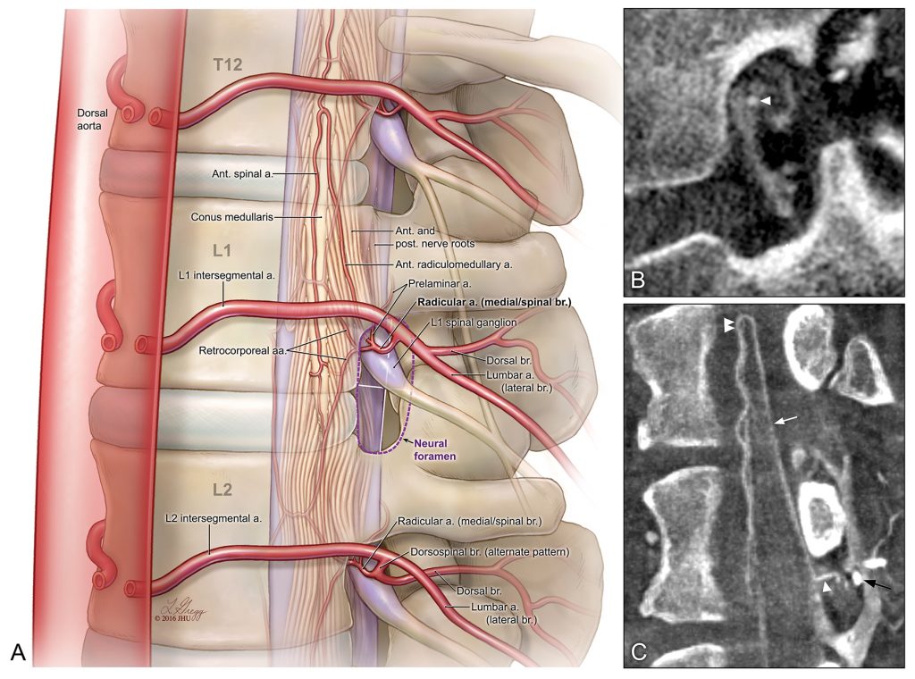
“Arterial anatomy of the neural foramen” Handbook of Clinical Neurology. Chapter 15, Gailloud P. © 2016 JHU, artist: Lydia Gregg.
It’s been a busy few weeks for faculty member, Lydia Gregg, MA, CMI, FAMI, who specializes in neurovascular spinal anatomy along with her collaborator, Dr. Philippe Gailloud. They work together to teach our Neuroanatomy for the Medical Illustrator course. The two of them have recently had 3 publications released:
First, the book, Handbook of Clinical Neurology, Vol 176, was published this month with Chapters written by both Ms. Gregg and Dr. Gailloud. Lydia was first author and illustrator of Chapter 3:
Gregg L & Gailloud P. “Neurovascular anatomy: Spine.” Handbook of Clinical Neurology. Edited by Hetts SW, Cooke DL, Vol 176. Elsevier 33-47.
https://www.sciencedirect.com/science/article/pii/B9780444640345000079.
Ms. Gregg also illustrated Chapter 15, by Dr. Gailloud:
Gailloud P. “Spinal vascular malformations: Angiographic evlauation and endovascular management.” Handbook of Clinical Neurology. Edited by Hetts SW, Cooke DL, Vol 176. Elsevier 267-304.
https://www.sciencedirect.com/science/article/pii/B9780444640345000134.
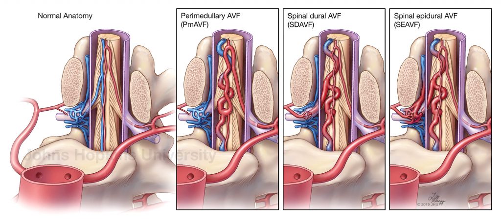
The Normal Anatomy and three main types of low-flow spinal arteriovenous fistulas (AVF): Perimedullary, Spinal dural, and Spinal epidural AVFs. © 2019 JHU, artist: Lydia Gregg.
Second, a new illustrated and co-authored paper in collaboration with Drs. Abderrahmane Hedjoudje, Olwen C. Murphy, and Carlos A. Pardo. The paper highlights three main types of low-flow spinal arteriovenous fistulas (AVF): perimedullary AVFs on the spinal cord surface, dural AVFs within the layers of the thecal sac, and epidural AVFs in the epidural space.
Hedjoudje A, Murphy OC, Gregg L, Pardo CA, Gailloud P. Spinal fistulas documented by contrast enhanced computed tomography during myelopathy workup: a lost opportunity. Neuroradiology. 2020 Nov 16:1-7.
https://link.springer.com/article/10.1007/s00234-020-02601-x.
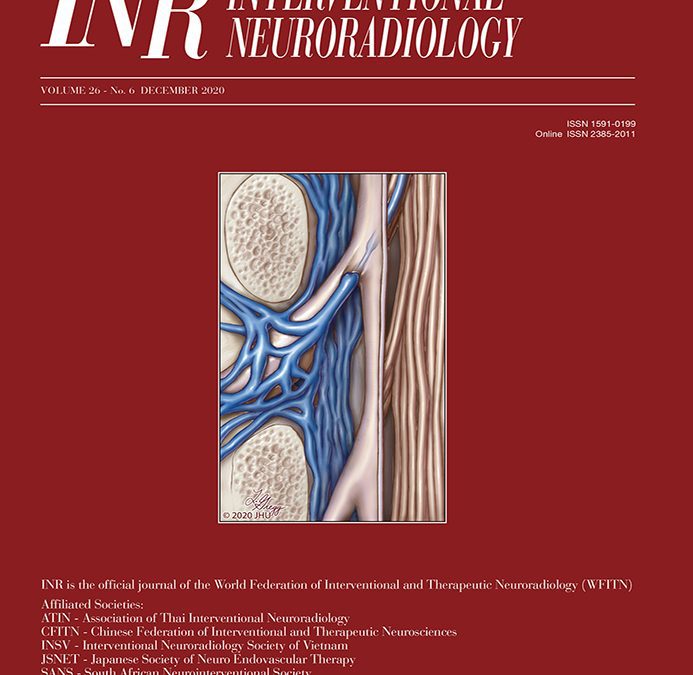
Interventional Neuroradiology, Vol 26, No 6; Dec 2020. Illustration of the antireflux mechanism of the spinal venous system. © 2020 JHU, artist: Lydia Gregg
Lastly, Ms. Gregg and Dr. Gailloud were also extremely honored to have their work featured on the cover of the journal, Interventional Neuroradiology, for the December 2020 issue, Vol 26 No 6. The Illustration is a depiction of the antireflux mechanism of the spinal venous system, a tight narrowing of the radiculomedullary vein at the dural crossing: Full article available here:
https://journals.sagepub.com/doi/full/10.1177/1591019920941309.
To view more of Lydia’s work as it’s published, follow her on twitter: @LydiaJGregg.