
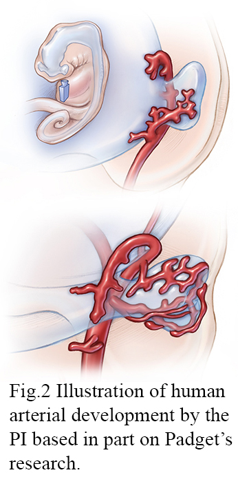
© JHU, Illustrated by Lydia Gregg based on art by Dorcas Hager Padget
© JHU
Congratulations to Lydia Gregg, MA, CMI, FAMI, on her receipt of a 2018 Catalyst Award, an intramural $75,000 grant!
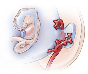
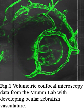
Lydia is an Associate Professor with a joint appointment in the Departments of Radiology and Art as Applied to Medicine. Her funded project entitled, Communicating Comparative Embryonic Arterial Development: An Interactive 3D Educational Tool Depicting Zebrafish and Human Ocular Arteries, seeks to visualize the development of major intracranial and ocular arteries by creating a comparative 3D educational tool that integrates human serial section data and zebrafish confocal microscopy data (Fig. 1). Developing this dynamic visualization will build on Lydia’s prior neurovascular embryology work (Fig. 2) 1-4 and experience illustrating, animating, and researching neurovascular anatomy 5-8.
References
- Gregg L, San Millán D, Orru’ E, Tamargo RJ, Gailloud P. Ventral and dorsal persistent primitive ophthalmic arteries. Operative Neurosurgery. 2015;12:141-152
- Gregg L, Gailloud P. The role of the primitive lateral basilovertebral anastomosis of padget in variations of the vertebrobasilar arterial system. Anatomical record (Hoboken, NJ: 2007). 2017;300:2025
- Gailloud P, Gregg L, Ruiz DS. Developmental anatomy, angiography, and clinical implications of orbital arterial variations involving the stapedial artery. Neuroimaging Clin N Am. 2009;19:169-179, Table of Contents
- Burger IM, Siclari F, Gregg L, Gailloud P. Bilateral segmental agenesis of the vertebrobasilar junction: Developmental and angiographic anatomy. AJNR Am J Neuroradiol. 2007;28:2017-2022
- Gregg L, Sorte D, Gailloud P. Intraforaminal location of thoracolumbar radicular arteries providing an anterior radiculomedullary artery using flat panel catheter angiotomography. American Journal of Neuroradiology. 2017;38:1054-1060
- Gregg L, Gailloud P. Transmedullary venous anastomoses: Anatomy and angiographic visualization using flat panel catheter angiotomography. American Journal of Neuroradiology. 2015
- Gailloud P, Gregg L, Pearl M, San Millan D. Ascending and descending thoracic vertebral arteries. American Journal of Neuroradiology. 2017;38:327-335
- Gailloud P, Gregg L, Katz Z, Myseros JS, Pearl MS. Duplicated origin of a radiculomedullary artery supplying a perimedullary arteriovenous fistula: Angiographic observation and developmental considerations. Anatomical science international. 2014;89:191-194\