Art as Applied to Medicine:
The First Program of its Kind…
At Johns Hopkins, medical illustration began with the arrival, in 1894, of Max Brödel, a young German artist from Leipzig, Germany. He had illustrated for Carl Ludwig in the famous Institute of Physiology at the University of Leipzig. There Brödel met American scientists who were studying under Ludwig. Later, one of these, anatomist Franklin P. Mall, urged young Brödel to join him at the new Johns Hopkins School of Medicine in Baltimore, Maryland.
Circumstances altered plans and upon arrival in Baltimore Brödel was quickly employed by Howard A. Kelly, Chief of Gynecology, as his illustrator for a two-volume textbook, Operative Gynecology. Other books followed, some with co-authors, on subjects as diverse as the vermiform appendix and diseases of the kidneys, ureters, and bladder. Aside from texts, journal articles, and monographs, Kelly and Brödel united in their efforts to advance the state of surgery and health care in America, especially in diseases of women. When time permitted, Brödel illustrated for other Hopkins physicians and surgeons, expanding his knowledge of anatomy, pathology, and physiology.
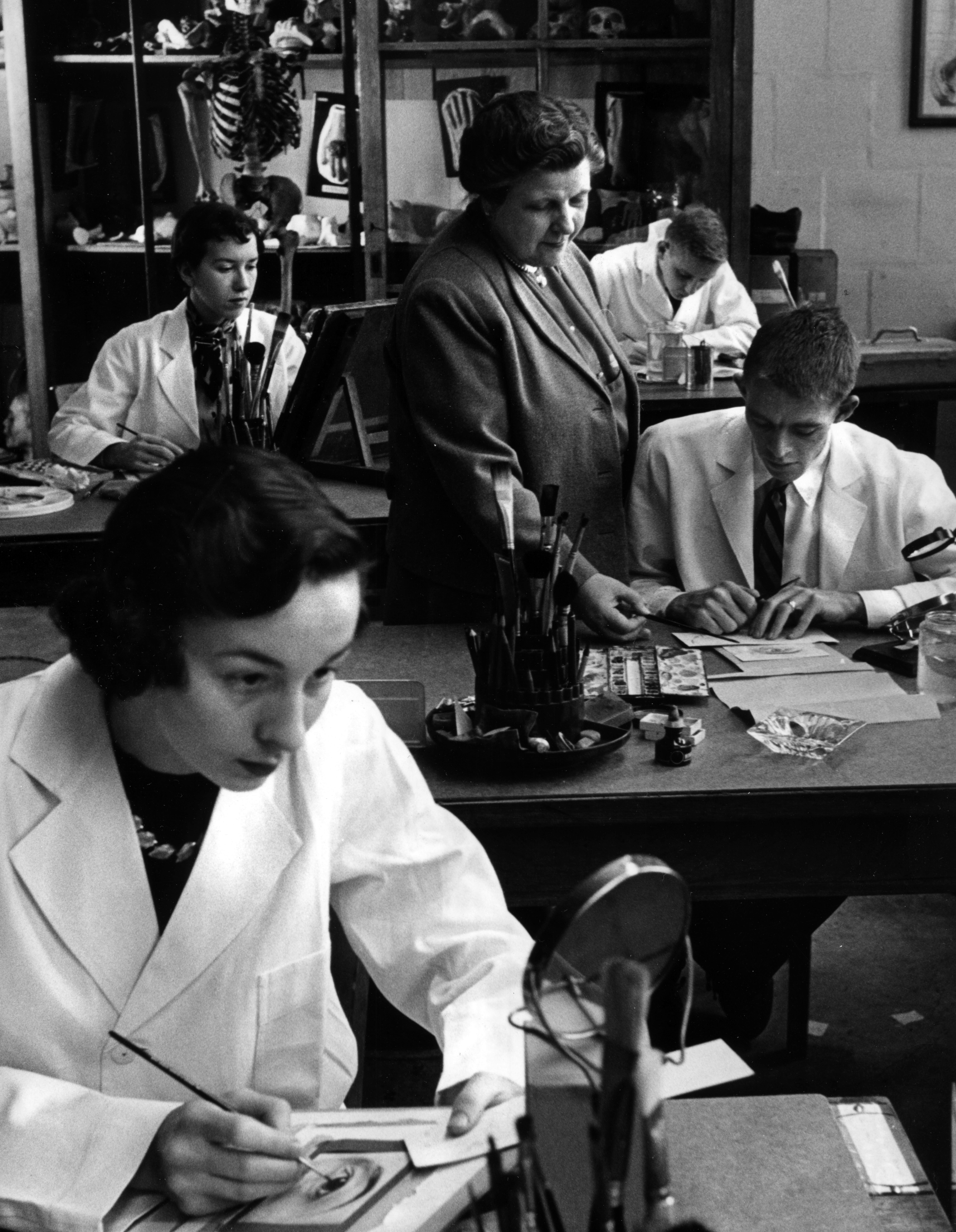
Medical illustration with all of its communication components and continually-evolving production technologies remains a vital discipline at JHMI. Faculty and students in this program are committed to continuing education in the medical sciences. We welcome the partnership with physicians, surgeons, and all other providers of medical and health care information to advance global medicine.
Our Centennial: Celebrating 100 Years
Art as Applied to Medicine, the first Department of its kind in the world, was endowed by Henry Walters in 1911 with Max Brödel as its first Director. This became the defining moment when the profession of Medical Illustration was established. Brödel arrived at Hopkins 17 years earlier in 1894 and from the beginning he realized that what he had embarked upon was something very new and different. In July of 2011 we presented a full day and evening of activities. We invite you to view the celebration of fellow medical illustrators, colleagues, friends and Hopkins alumns in a fun and informative reunion. Here at Hopkins we are on the dawn of exciting scientific, medical, and communication technologies but still teaching the “art” of our profession. Our one-day celebration not only takes a look at the historical past but address the advances along the way and try to project the future of the profession and the skills all of the graduate programs we will be teaching in the next 100 years.A catalog of A Century of Medical Illustration is now available as a print-on-demand book. This 358 page, hardbound, full-color catalog features the artwork of the Johns Hopkins Centennial Exhibition, celebrating 100 years of teaching excellence in medical illustration. It is available online at Blurb.com. Content was compiled by the Centennial Committee at Johns Hopkins and the book was designed by Zina Deretsky. No proceeds are being made on its sale.
Related Links:
- Purchase a Kidney Stone print.
- JHU Gazette, A Century of Medical Illustration
- Read about the exhibition on the JH Pathology Blog
- View photos taken at the Centennial Program and Evening Celebration…
- View a PDF of the Centennial Program.
In Memorium
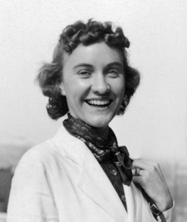
Betty J. Blumenthal
1917 - 2006
Elizabeth "Betty" Blumenthal was a medical sculptor and member of the faculty of the Department of Art as Applied to Medicine for over 53 years. In her professional work, Betty designed and fabricated sub-dermal implants for patients with birth defects, deforming injuries, or recovering from cancer surgery. For more than three decades, notable plastic surgeons sought her skills for their patients.
Betty was a 1933 graduate of Western High School in Baltimore. She studied drawing at the Maryland Institute College of Art and later studied at the Schuler School of Fine Art in Baltimore. 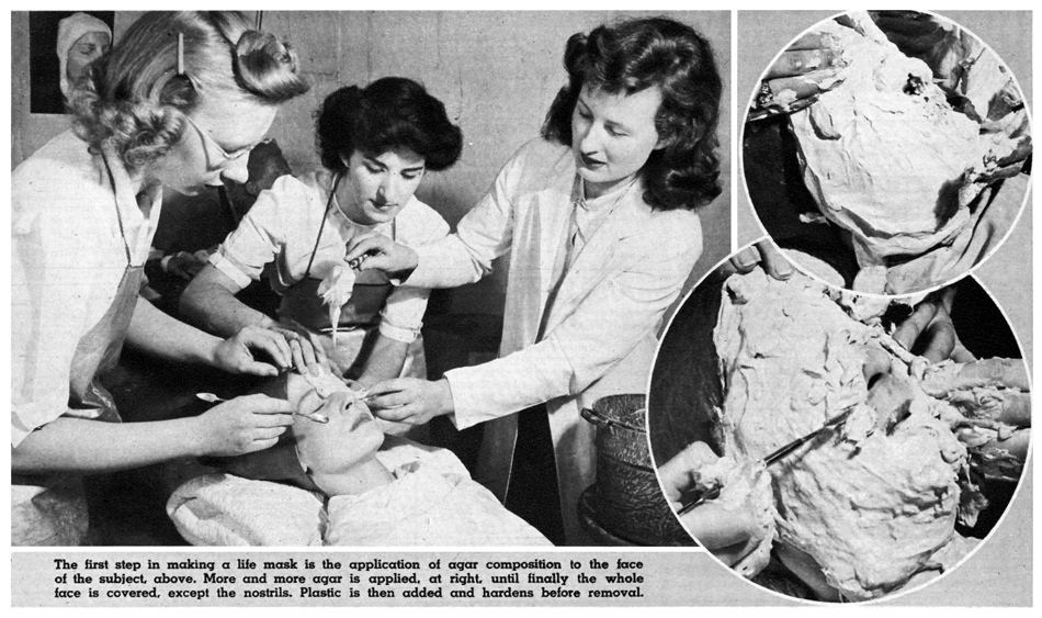 In early 1943 when Ranice Crosby became the Director of the Department of Art as Applied to Medicine at Hopkins, Betty was director of the Photography unit at Johns Hopkins Hospital. They had been working together on an obstetrics book for Dr. Nicholson Eastman, so when Ranice decided to add photography to the curriculum, Betty was an excellent choice to teach the course. She taught photography for several years, and in 1944 added moulage (molding and casting body parts) to her instruction duties. Her love for medical sculpture came from her training in medical art at the University of Maryland under Carl Dame Clark. In 1946 The Sunday Sun Magazine (of The Baltimore Sun newspaper) had an article on the department at Hopkins and quoted Betty as saying "when she got a chance to work at the Hopkins, she knew that dreams do, sometimes, come true."
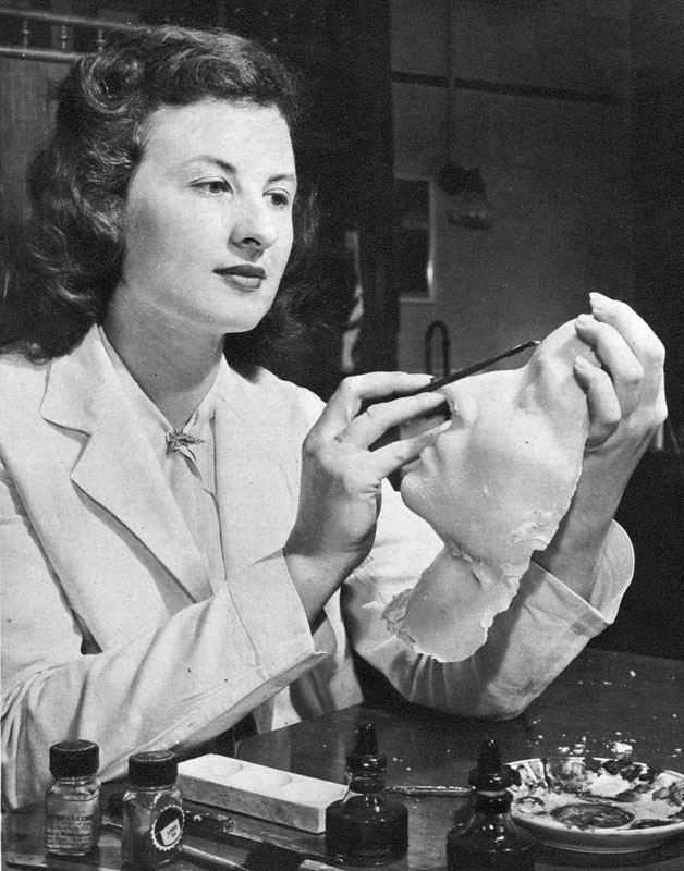
Her knowledge of prosthetics and medical sculpture has been passed on to a lengthy legacy of medical illustrators. Students, including Mary Ann Shumate, Gerald Hodge, and John Cody in the 40's; Al Teoli, Neil Hardy, and Herb Smith in the 50's; and Tim Hengst, Mark Miller, and Juan Garcia in the 70's 80's and 90's, all gained knowledge from her instruction. Her skills in patient casting and prosthesis sculpting made her a valuable asset in our department. Her teaching was a precursor to the Facial Prosthetics clinic operating in our department today. Always poised and smiling, Betty was a lovely presence in our department for many years.
Annette S. Burgess
1899 - 1962
Annette Smith Burgess came to Johns Hopkins in 1923 and studied with the founding director of the Department of Art as Applied to Medicine, Max Brödel for three years. Under his rigorous tutelage, she perfected her technique to become one of his greatest pupils. In 1926 at the behest of Dr. William H. Wilmer, she became a full-time Ophthalmic Illustrator with the Wilmer Institute.
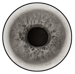
Annette Burgess painted and drew practically all of the illustrations that appeared in the various medical publications of the Wilmer Institute. She became the foremost painter of the ocular fundus and also a world-renowned ophthalmic artist. Annette Burgess’ work was marked by her accuracy of observation, her meticulous attention to detail, her absolute honesty in depicting pathological lesions, and her uncompromising refusal to include any lesion not actually present in the particular eye under examination. Her honesty, integrity, and loyalty were highly regarded by her colleagues, admirers, and friends. She served the Institute until her retirement in 1962.