
Faculty members Jennifer Fairman and Lydia Gregg received multiple awards in the Biocommunications Association’s 2025 Bioimages Exhibit. You can view the entire exhibit here, and read below about our faculty’s awards:
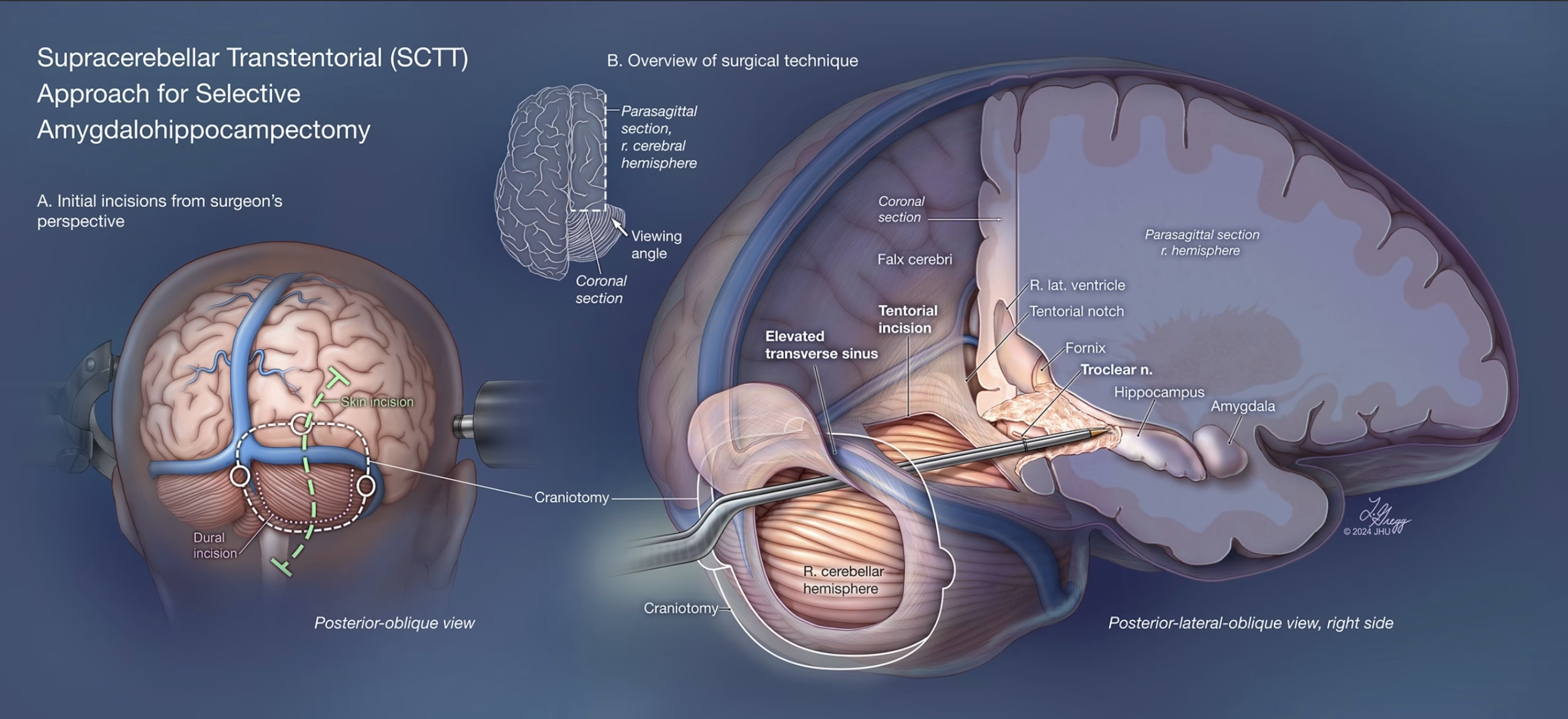
Supracerebellar Transtentorial Approach for Selective Amygdalohippocampectomy
Premier Award, Illustration & Graphic Media Division | Medical Illustration
Lydia Gregg, MA, CMI, FAMI
This illustration summarizes the supracerebellar transtentorial approach for selective amygdalohippocampectomy. This posterior approach through a tentorial incision involves elevating the transverse sinus to access temporal lobe structures such as the hippocampus and amygdala.
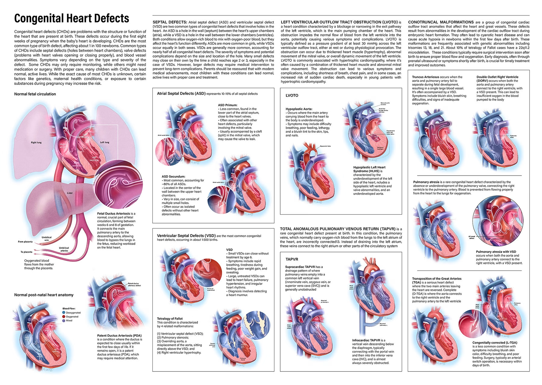
Congenital Heart Defects
BCA Medical Education Award/ Illustration and Graphic Media Division; Image of Merit, Illustration & Graphic Media Division | Graphic Design Media
Jennifer Fairman, MA, MPS, CMI, FAMI
This illustration was awarded for the best medical teaching aid in the Illustration and Graphic Media Division. Congenital heart defects (CHDs) are structural problems with the heart present at birth, affecting approximately 1 in every 100 infants born in the United States. These defects occur during the first eight weeks of pregnancy when the heart is developing. CHDs can involve different parts of the heart, such as the atria and ventricles (the chambers), valves, and major blood vessels. They can lead to abnormal blood flow, either too much or too little, through the lungs or the body, which can result in life-threatening conditions if left untreated. This poster showcases the variety of CHDs and their unique anatomical configurations.
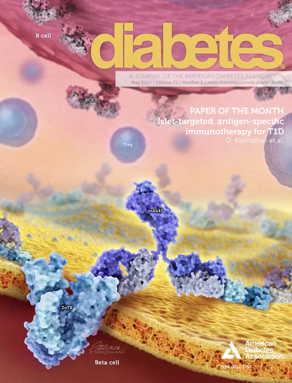
Islet-Targeted, Antigen-Specific Immunotherapy for T1D
Image of Distinction, Illustration & Graphic Media Division | Graphic Design Media
Jennifer Fairman, MA, MPS, CMI, FAMI
This image was created to visually summarize and highlight the key scientific findings and recent innovations presented in the ‘Paper of the Month’ of Diabetes (Diabetes 2024 May 1;73(5):806-818. doi: 10.2337/db23-0568). Its purpose is to provide a concise and accessible overview of the article’s core concepts, specifically geared for physicians, healthcare professionals, and other members of the American Diabetes Association’s journal readership to quickly grasp the study’s significance and applications to the prevention of an autoimmune response and to reverse the onset of T1D.
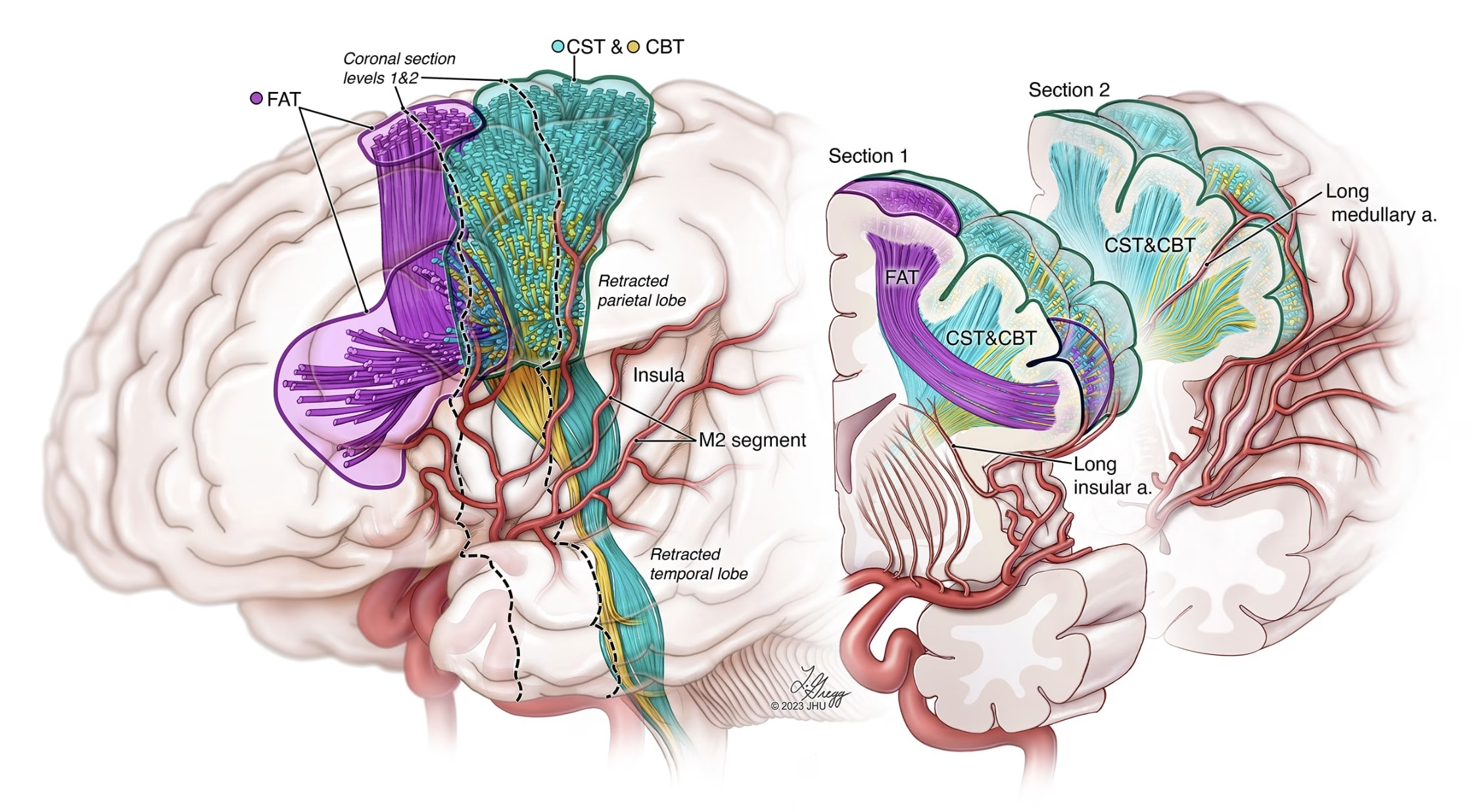
Motor Risks of Insular Epilepsy Surgery
Image of Distinction, Illustration & Graphic Media Division | Medical Illustration
Lydia Gregg, MA, CMI, FAMI
This illustration depicts motor structures of the brain that may be damaged during insular surgery including the FAT (frontal aslant tract), CST (corticospinal tract), and CBT (corticobulbar tract) pathways. The M2 segments of the MCA branches cover and supply the insula before they extend over the cerebral hemispheres. The two coronal sections on the right show how the long insular artery and the long medullary artery supply the corticospinal and corticobulbar tracts. These crucial branches must remain intact during surgery.
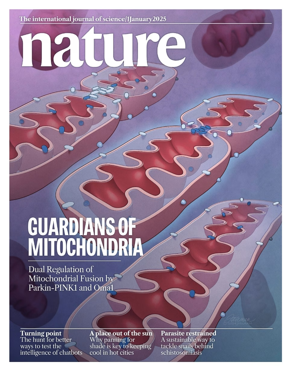
Guardians of Mitochondria: Dual Mitochondrial Fusion by Parkin-PINK1 and Oma1
Image of Merit, Illustration & Graphic Media Division | Graphic Design Media
Jennifer Fairman, MA, MPS, CMI, FAMI
This proposed cover illustrates the regulation of mitochondrial fusion. Mitochondria have two membranes, and both must work together to safeguard proper fusion, but they use different processes and machinery. Mitochondrial stress pathways help protect mitochondria from overfusion and damage, though their role under normal conditions is not well understood. This study, using mice with different gene mutations, highlights two stress-response systems, Parkin and Oma1, that work together to protect mitochondrial structure and DNA. They help mitochondria fuse correctly, with assistance from two proteins: Mfn1 in the outer membrane and Opa1 in the inner membrane. Losing both Parkin and Oma1 leads to small body size, low activity, early death, and mitochondrial issues. These results show that Parkin and Oma1 are crucial for controlling mitochondrial fusion, even without external stress.