
Save the Date — Friday, April 19, 2013
The Lecture:
Friday, April 19, 2013
3:30 P.M.
Mountcastle Auditorium
Preclinical Teaching Building
725 North Wolfe Street
The Johns Hopkins University
School of Medicine
Baltimore
Reception to follow.
Members of the scientific and art communities are cordially invited to attend.
Graham Johnson, Ph.D.
 Graham Johnson has specialized in visualizing molecular and cellular biology since graduating from Johns Hopkins in 1997 with a masters degree in Medical and Biological Illustration. He illustrated both editions of the textbook Cell Biology by Pollard & Earnshaw as a coauthor and has created thousands of scientific visuals ranging from journal covers to pedagogic animations. Graham’s recent biophysics PhD work in the Molecular Graphics Lab at The Scripps Research Institute focused primarily on developing algorithms to enable scientists and illustrators to generate, simulate, and visualize molecular models of cells. Now with his own lab at The University of California San Francisco, as a QB3 Faculty Fellow, he continues to work with programmers to develop software that can interoperate the computational tools of science and art.
http://grahamj.com
UCSF Lab: http://mesoscope.org
The Samson Feldman Visiting Scholar
In Art as Applied to Medicine
Rossetta A. and Sadie B. Feldman, sisters of Samson Feldman, have established a visiting lectureship to honor his life as an artist and lifelong patron of the arts. Samson Feldman always believed in the everpresent relationship of art and science. It is appropriate to designate the field of art as related to medicine as the topic to which the lectures will be addressed. The lectures are to be selected from distinguished scholars in visual communications with the purpose of presenting contemporary views pertaining to medical art. The selection of lecturers will be made by a committee representing The Department of Art as Applied to Medicine.
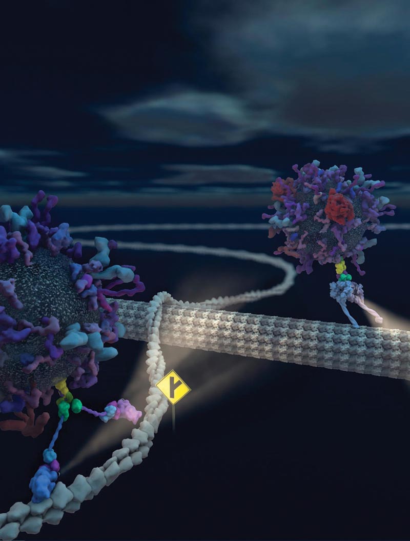 Illustration: Cell Cargo Transport Junction
Motor proteins “walk” along cytoskeletal filaments that web throughout the interiors of most of the cells. The cytoskeletal filaments support the structure and shape of the cell, but also serve as highways to transport cargo from one part of the cell to another efficiently. This image shows the trafficking of cargo vesicles along actin filaments and microtubles, with the intersection symbolizing the transition between cytoskeletal elements. In this issue of Neuron, the authors Heisler et al. offer insight into how the switch between these transport systems can occur, demonstrating that muskelin guides GABAA receptors across cytoskeletal tracks by connecting to both myosin VI and dynein motor complexes. Vesicle models were generated with the mesoscale modeling software autoFill developed by the artist Graham Johnson in Art Olson’s Molecular Graphics Lab (MGL) at the Scripps Research Institute. He then created and rendered the cytoskeletal and motor protein models with ePMVC4D, also developed by Ludovic Autin and Johnson at MGL and UCSF.